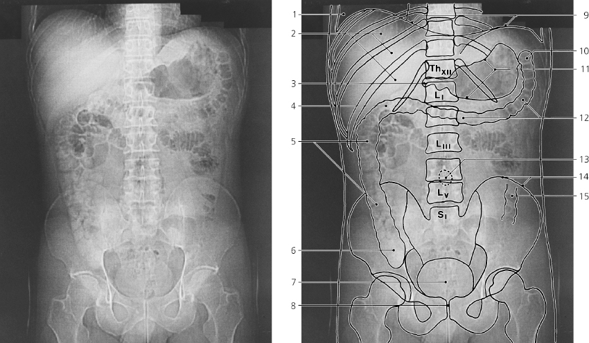
The gastro-intestinal tract is outlined by its natural gas content
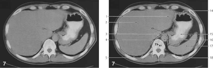
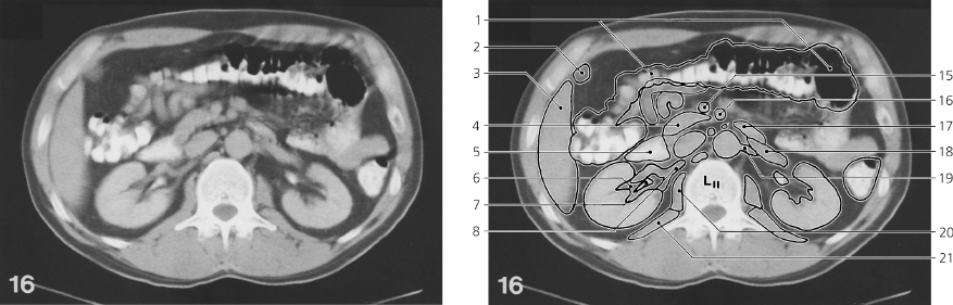
Diaphragm
Costodiaphragmatic sulcus
Mediastinodiaphragmatic sulcus
Lower border of liver
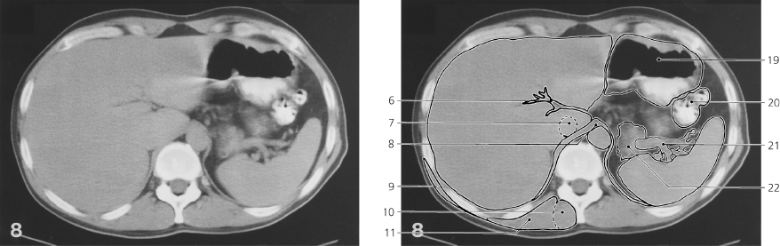
Hepatic flexure of colon
Duodenal cap (radiology term)
Ascending colon
Upper pole of right kidney
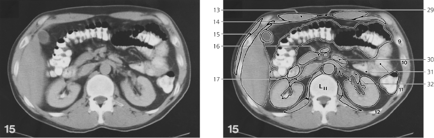
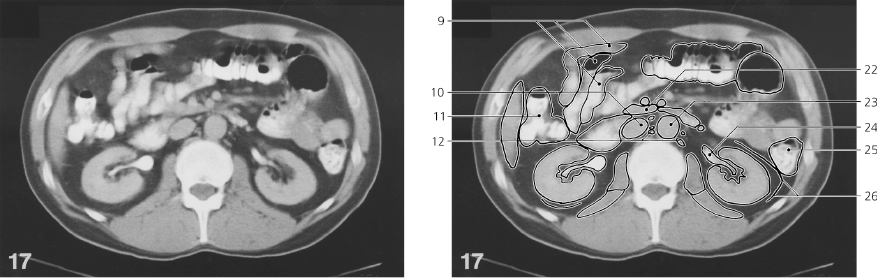
Psoas major (lateral contour)
Cecum
Lower border of spleen
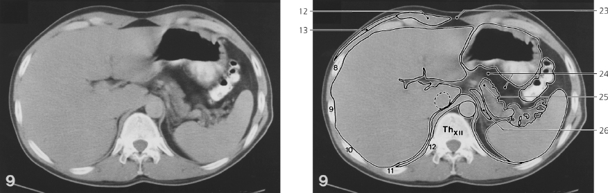
Splenic flexure of colon
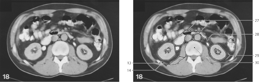
13: 12th rib
Stomach
Descending colon
Jejunum
Lower pole of left kidney
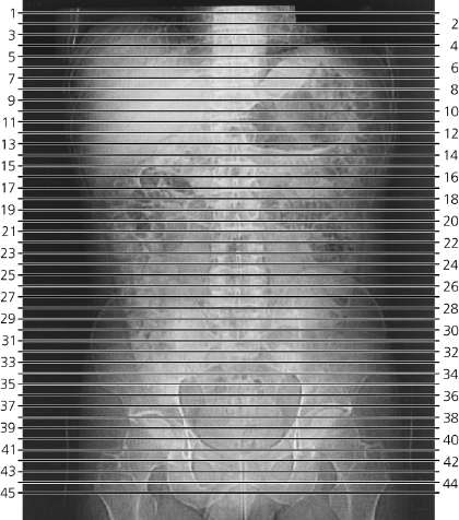
Scout view
Costodiaphragmatic sulcus
Liver
Duodenal cap
Hepatic flexure of colon
Ascending colon
Cecum
Urinary bladder
Symphysis pubis
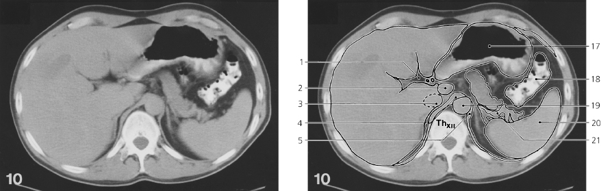
Diaphragm
Splenic flexure of colon
Curvatures of stomach
Transverse colon
Position of umbilicus
Iliac crest
Descending colon
Scout view
Lines #1–45 indicate position of sections in the following CT series. Consecutive sections, 10 mm thick. The gastrointestinal tract is outlined by peroral contrast medium. The urinary tract is outlined by excretion of intravenous watersoluble contrast medium. Residues of contrast from an earlier lymphography are present in some iliac and lumbar lymph nodes.
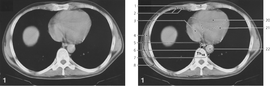
Abdomen, axial CT
Scout view on opposite page
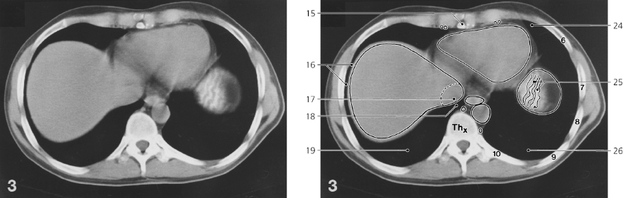
Body of sternum
Calcified costal cartilage
Right atrium
Serratus anterior
Esophagus
Azygos vein
Hemiazygos vein
Latissimus dorsi
Internal thoracic artery and vein
Diaphragm
Right lobe of liver
Inferior caval vein
Iliocostalis thoracis, and longissimus thoracis
Transversospinal muscles
Xiphoid process
Costodiaphragmatic groove
Inferior caval vein
Phrenico-mediastinal groove
Lower lobe of right lung
Right ventricle
Left ventricle
Thoracic aorta
Spinal cord
Lingula of left lung
Rugae in fundus of stomach
Lower lobe of left lung Ribs are numbered.
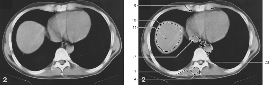
Abdomen, axial CT
Scout view on page 404
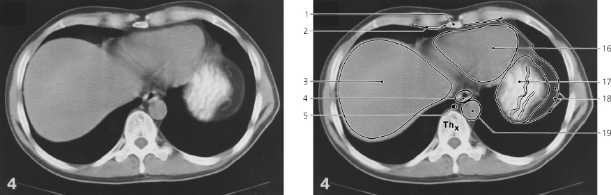
Xiphoid process
Transversus thoracis
Right lobe of liver
Esophagus
Azygos vein
Costal cartilage
Costo-diaphragmatic groove with inferior margin of right lung
Serratus anterior
Inferior caval vein
Latissimus dorsi
Phrenico-mediastinal groove
Thoracolumbar fascia
Rectus abdominis
Obliquus externus abdominis
Caudate lobe of liver
Heart
Fundus of stomach with rugae
Parietal pleura, diaphragm, and parietal peritoneum
Thoracic aorta
Apex of heart
Esophagus, abdominal part
Left lobe of liver
Oblique fissure of left lung
Fundus of stomach with air and barium
Cardia
Spleen Ribs are numbered.
Abdomen, axial CT
Scout view on page 404
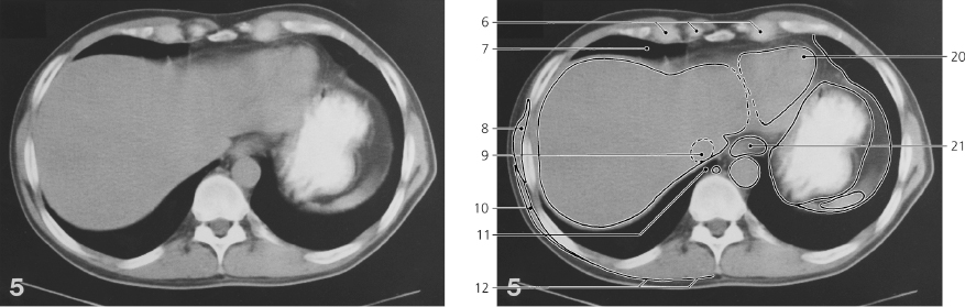
Left lobe of liver
Right lobe of liver
Caudate lobe of liver
Lumbar part of diaphragm
Inferior margin of left lung
Porta hepatis
Inferior caval vein
Right crus of diaphragm
Latissimus dorsi
Transversospinal muscles
Iliocostalis and longissimus
Rectus abdominis
Obliquus externus abdominis
Rugae in fundus of stomach
Body of stomach
Thoracic aorta
Spleen
Inferior margin of left lung
Air in body of stomach
Splenic flexure of colon
Splenic vessels
Tail of pancreas
Linea alba
Omental bursa with surrounding peritoneal fat
Body of pancreas
Splenic artery Ribs are numbered.
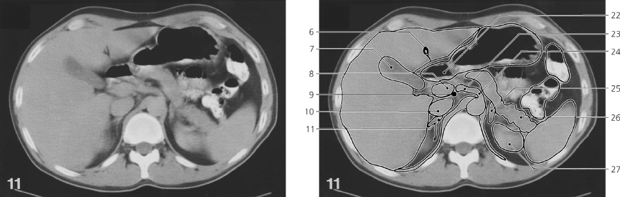
Abdomen, axial CT
Scout view on page 404
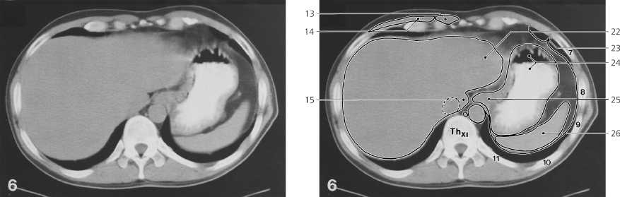
Porta hepatis
Portal vein
Inferior caval vein
Right crus of diaphragm
Left crus of diaphragm
Lig. teres hepatis
Gall bladder
Portal vein
Bile duct (choledochus)
Inferior caval vein
Right suprarenal gland
Left lobe of liver
Wall of gall bladder
Head of pancreas
Superior part of duodenum
Upper pole of right kidney
Body of stomach
Splenic flexure of colon
Abdominal aorta
Spleen
Splenic vessels
Duodenal “cap” (bulbus)
Common hepatic artery
Celiac trunk
Left suprarenal gland
Tail of pancreas
Upper pole of left kidney
Portal vein behind pancreas
Transverse colon
Body of pancreas
Jejunum with air and barium
Descending colon
Diaphragm
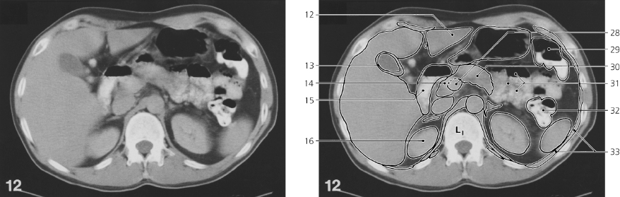
Abdomen, axial CT
Scout view on page 404
Left lobe of liver
Hepatic flexure of colon
Superior part of duodenum
Head of pancreas
Right suprarenal gland
Right crus of diaphragm
Left crus of diaphragm
Fundus of gall bladder
Inferior caval vein
Descending part of duodenum
Right kidney
Quadratus lumborum
Rectus abdominis
Transversus abdominis
Obliquus externus abdominis
Uncinate process of pancreas
Right renal vein
Portal vein
Splenic vein
Superior mesenteric artery
Left suprarenal gland
Descending colon
Sinus renalis
Transverse colon
Superior mesenteric vein
Duodenojejunal flexure
Superior mesenteric artery
Abdominal aorta
Linea alba
Ascending part of duodenum
Jejunum
Left renal vein
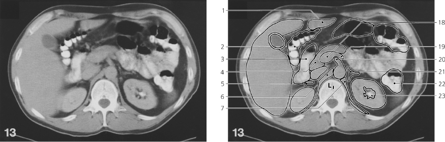
Abdomen, axial CT
Only gold members can continue reading. Log In or Register to continue