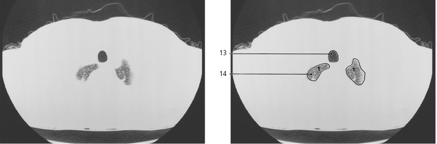
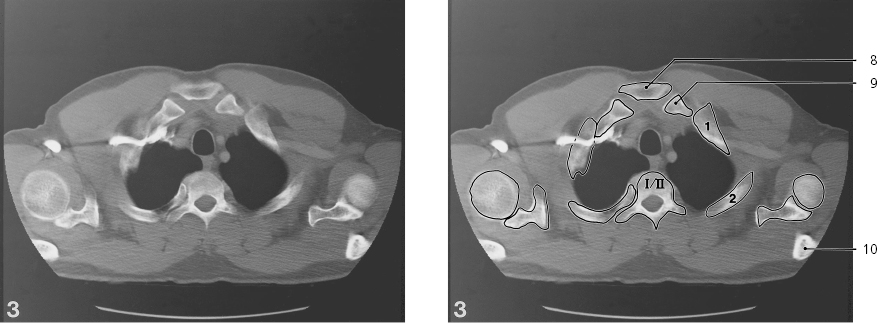
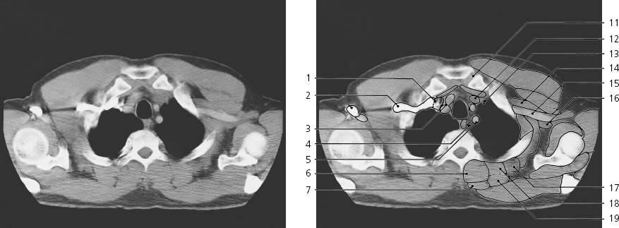
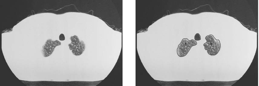
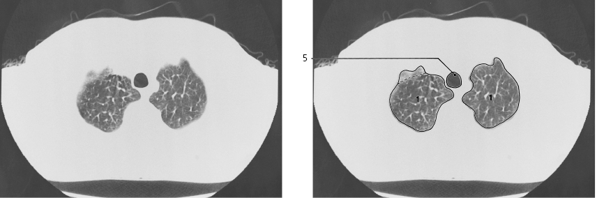
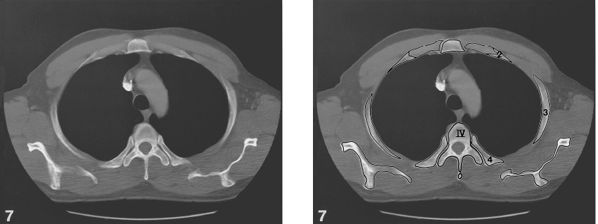
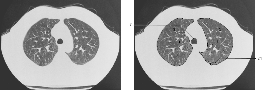
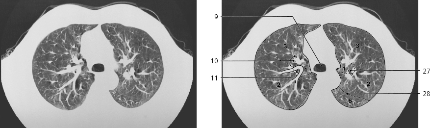
Lines #1–32 indicate positions of axial sections in the following CT series. All sections are 5 mm thick and are spaced by 5–20 mm. Each section is displayed with bone settings (above), soft tissue settings (middle), and lung settings (below). Arms are raised above head. Intravenous contrast was given in the right cubital vein. Vertebrae are numbered with romans and costae with arabics on the bone image. Lung segments are numbered with arabics on the lung image.
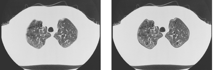
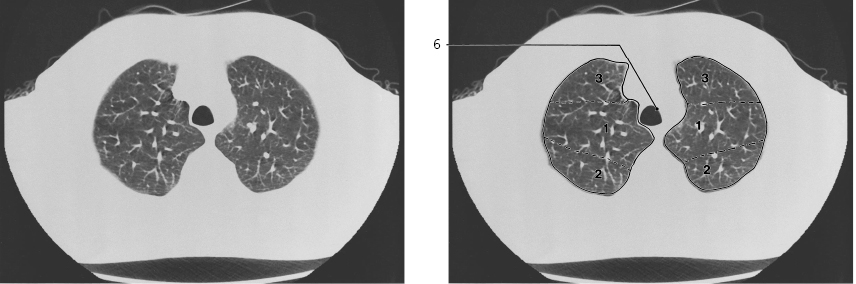
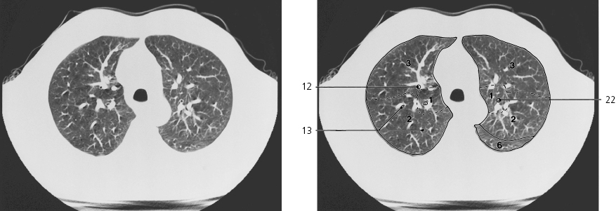
Right lung segments
Superior lobe:
# 1: Apical segment
# 2: Posterior segment
# 3: Anterior segment
Middle lobe:
# 4: Lateral segment
# 5: Medial segment
Inferior lobe:
# 6: Superior segment
# 7: Medial basal segment
# 8: Anterior basal segment
# 9: Lateral basal segment
#10: Posterior basal segment
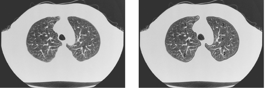
Left lung segments
Superior lobe:
# 1: Apical segment
# 2: Posterior segment
# 3: Anterior segment
# 4: Superior lingular segment
# 5: Inferior lingular segment
Inferior Lobe:
# 6: Superior segment
# 7: Medial basal segment
# 8: Anterior basal segment
# 9: Lateral basal segment
#10: Posterior basal segment
#1 and #2 of left lung usually arise from a common apicoposterior segmental bronchus. Note that the diameter of bronchi appear very narrow in the lung image due to the partial volume effect in CT imaging. Arrows ←, → and ↔ in the legends indicate that a structure can be seen on a previous or following section, or both.
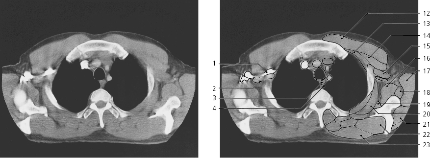
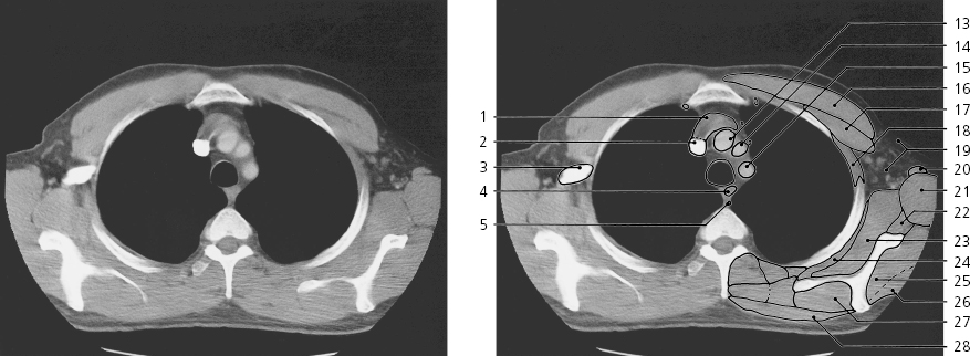
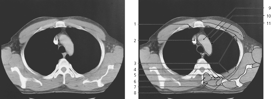
Thorax, axial CT (scout view on previous page)
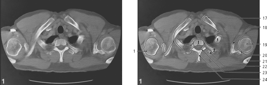
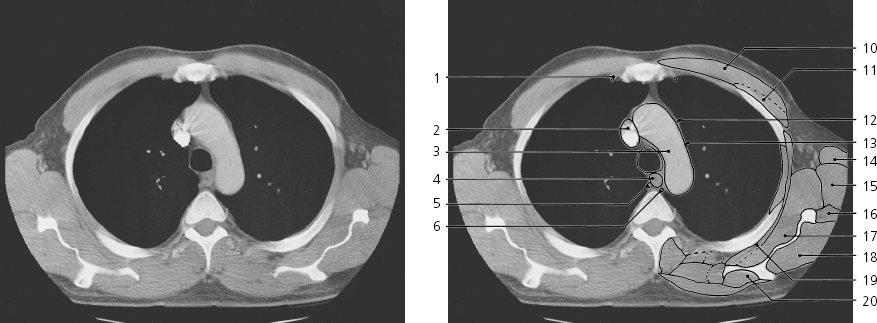
Greater tubercle of humerus →
Anterior jugular vein
Right common carotid artery →
Internal jugular vein (with contrast)
Right subclavian artery →
Axillary vein (with contrast) →
Axillary artery →
Lower pole of thyroid lobe
Esophagus →
Left internal carotid artery →
Lymph node
Scalenus anterior muscle →
Left subclavian artery →
Rhomboideus →
Trachea →
Apex of lung →
Sternal end of clavicle →
Coracoid process →
Head of humerus →
Glenoid cavity →
Acromion →
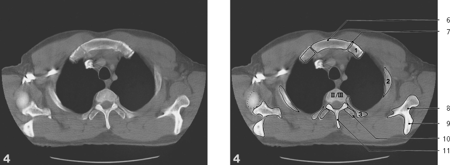
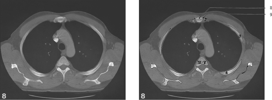
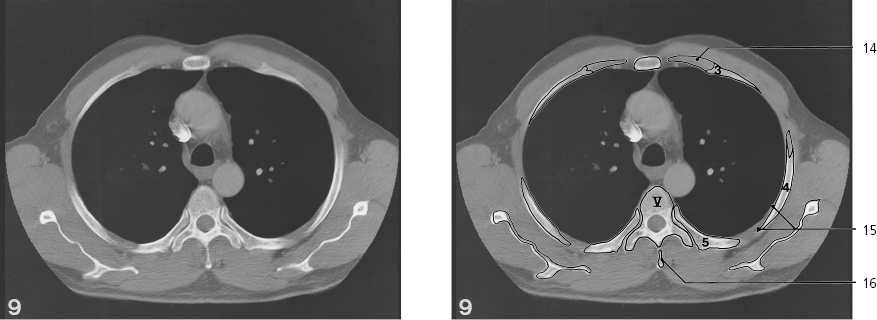
Transverse process of Th II
Lamina of vertebral arch
Spinous process of Th I
Sternocleidomastoideus, sternal head →
Sternothyroideus and sternohyoideus →
Pectoralis major →
Subclavius muscle →
Pectoralis minor →
Axillary fossa →
Teres major →
Biceps brachii, short head
Subscapularis muscle →
Iliocostalis cervicis →
Supraspinatus →
Trapezius →
Longissimus →
Levator scapulae →
Transversospinal muscles →
Thorax, axial CT (scout view on page 351)
Processus coracoideus ←
Greater tubercle of humerus ←
Right common carotid artery ←
Subclavian and internal jugular vein, confluence ↔
Internal thoracic artery →
Scalenus anterior (insertion) ←
Axillary vein (with contrast) ↔
Axillary artery ↔
Right subclavian artery ←
Left common carotid artery ↔
Esophagus ↔
Left subclavian artery ↔
Trachea ↔
Apex of lung ←
Sternal end of clavicle ↔
Head of humerus ↔
Glenohumeral joint ↔
Sternocleidomastoideus, sternal head ↔
Interclavicular ligament
Articular disc of sternoclavicular joint →
Sternohyoid and sternothyroid muscles ↔
Subclavius muscle ←
Pectoralis major ↔
Pectoralis minor ↔
Teres major ↔
Subscapularis ↔
Serratus anterior →
Supraspinatus ↔
Trapezius ↔
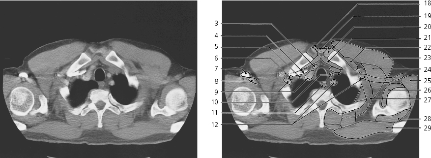
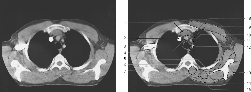
Thorax, axial CT (scout view on page 351)
Confluence of subclavian and internal jugular veins ←
Axillary vein (with contrast) ↔
Division of brachiocephalic trunk →
Thoracic duct →
Left subclavian artery ↔
Transversospinal muscles ↔
Rhomboideus ↔
Manubrium of sternum →
Sternal end of clavicle ←
Acromion ←
Articular disc of sternoclavicular joint ←
Left brachiocephalic vein ←
Internal thoracic artery ↔
Right axillary vein ↔
Right axillary artery ↔
Omohyoideus, inferior belly ←
Iliocostalis ↔
Longissimus ↔
Levator scapulae ↔
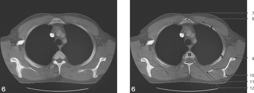
Thorax, axial CT (scout view on page 351)
Right axillary vein with contrast ↔
Axillary artery ←
Esophagus ↔
Thoracic duct ↔
Trachea ↔
Manubrium of sternum ↔
Synchondrosis of first rib
Neck of scapula
Spine of scapula →
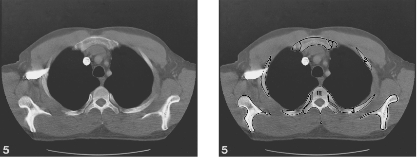
Transverse process of Th III
Spinous process of Th II
Pectoralis major ↔
Intercostal muscles ↔
Pectoralis minor ↔
Left axillary vein ←
Subscapularis ↔
Teres major ↔
Teres minor →
Omohyoideus, inferior belly ←
Serratus anterior ↔
Infraspinatus →
Supraspinatus ↔
Trapezius ↔
Thorax, axial CT (scout view on page 351)
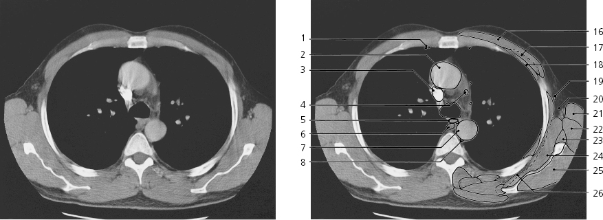
Left brachiocephalic vein ↔
Right brachiocephalic vein ↔
Right axillary vein →
Brachiocephalic trunk ↔
Iliocostalis ↔
Longissimus ↔
Transversospinal muscles ↔
Internal thoracic artery ↔
Left phrenic nerve →
Left vagus nerve →
Axillary fossa with nerves, vessels and lymph nodes ↔
Left subclavian artery ←
Deltoideus ↔
Levator scapulae ↔
Rhomboideus ↔
Thorax, axial CT (scout view on page 351)
Left brachiocephalic vein ↔
Right brachiocephalic vein ↔
Right axillary vein ←
Esophagus ↔
Thoracic duct ↔
Trachea ↔
Manubrium of sternum ↔
Costal cartilage ↔
Lateral margin of scapula
Spine of scapula ↔
Medial margin of scapula
Zygapophyseal joint Th III-IV
Brachiocephalic trunk ←
Left common carotid artery ←
Left subclavian artery ←
Pectoralis major ↔
Pectoralis minor ↔
Intercostal muscles ↔
Axillary fossa with nerves, vessels and lymph nodes ↔
Latissimus dorsi →
Teres major ↔
Teres minor ↔
Subscapularis ↔
Serratus anterior ↔
Infraspinatus ↔
Deltoideus ←
Supraspinatus ↔
Trapezius ↔
Thorax, axial CT (scout view on page 351)
Internal thoracic artery and vein ↔
Confluence of right and left brachiocephalic veins ←
Intercostal muscles ↔
Iliocostalis ↔
Longissimus ↔
Transversospinal muscles ↔
Rhomboideus ↔
Levator scapulae ↔
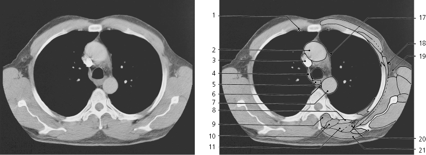
Aortic arch →
Left phrenic nerve ↔
Left vagus nerve ↔
Thorax, axial CT (scout view on page 351)
Internal thoracic artery and vein ↔
Superior caval vein →
Aortic arch ←
Esophagus ↔
Azygos vein (right superior intercostal vein) →
Thoracic duct ↔
Trachea ↔
Body of sternum →
Sternocostal joint of second rib
Pectoralis major ↔
Pectoralis minor ↔
Left phrenic nerve ↔
Left vagus nerve ↔
Latissimus dorsi ↔
Teres major ↔
Teres minor ↔
Subscapularis ↔
Infraspinatus ↔
Serratus anterior ↔
Supraspinatus ↔
Oblique fissure of left lung →
Thorax, axial CT (scout view on page 351)
Internal thoracic artery and vein ↔
Ascending aorta →
Superior caval vein ↔
Azygos vein (arch) →
Esophagus ↔
Thoracic duct ↔
Descending aorta →
Iliocostalis
Transversospinal muscles ↔
Longissimus ↔
Rhomboideus ↔
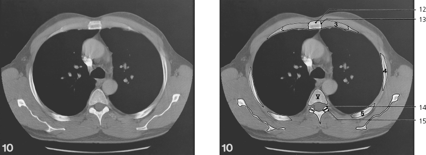
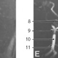
Branch of anterior segmental bronchus B III →
Branches of posterior segmental bronchus B II
Costal cartilage ↔
Costal sulcus
Spinous process of Th IV
Left phrenic nerve ↔
Lateral thoracic artery →
Axillary fossa ↔
Levator scapulae ↔
Trapezius ↔
Apical segmental bronchus B I
Thorax, axial CT (scout view on page 351)
Only gold members can continue reading. Log In or Register to continue