(up to tens of mT) to boost the sample magnetization and a much smaller detection field  (~μT) in which the magnetization precesses. For the DC effect,
  was perpendicular to the current dipole and
  was turned off non-adiabatically. For the AC effect,
  was parallel to the current dipole and
  turned off adiabatically.
  was set to 1.93 μT corresponding to a Larmor frequency of 82 Hz which matches the main frequency band of the evoked N20. A
  of 5 mT was applied in both cases.
In Fig. 1 the results of the DC and AC effect are shown. For the DC effect the amplitude spectra of of the difference of the time domain signals (phantom on–phantom off) are shown. The subtraction is necessary to reveal the minute effect on the NMR line-shape. The amplitude of the residual signal scales with the applied current dipole moment Q and we can infer a resolution limit of ~200 nAm.
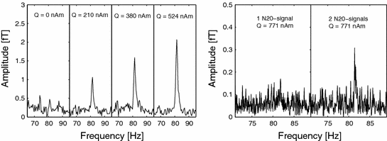
Fig. 1
Left Results for the DC effect phantom measurements (4,000 averages, total measurement time: 4 h 27 min). The residual signal scales with Q and a resolution limit of ~200 nAm was obtained. Right Results for the AC effect phantom measurements (1,000 averages, total measurement time: 4 h 10 min). Two consecutive N20 signals with Q = 771 nAm induced a reliable resonant signal in contrast to the application of a single N20 trace. The resolution limit was estimated to be ~1 μAm
For the AC effect, applying two physiological N20 signals consecutively with Q = 771 nAm clearly induces an NMR signal at 82 Hz. For a single N20 signal with the same Q a signal cannot be identified reliably and we conclude, that a resolution limit is likely to be about 1 μAm for a single N20 signal. Comparing these resolution limits to the ECDs evoked by electrostimulation of the median nerve it is evident that an increase of the signal to noise ratio (SNR) of at least 4 is necessary to observe neuronal currents based on the DC effect. The AC effect requires an increase in SNR of at least 67 and appears to be more difficult to exploit.
It should be noted that the quoted resolution limits represent lower bounds. The physiological ECDs are in fact deeper than current dipole in the phantom and the NMR relaxation times are significantly longer, in particular for the study regarding the AC effect, than the physiologically observed values for T 2 of 106 ms (gray matter) and 79 ms (white matter) (Zotev et al. 2009). An increase of the noise by a factor of 3 would arise if the excessively long measurement time of about 4.5 h was reduced to 30 min. The final minimum resolution limits are then 600 nAm and 3 μAm for the DC and AC effects, respectively.
3 Imaging Below 1 kHz
For the potential use of the AC effect in ULF MRI, initial 2D MRI experiments were performed inside a custom designed magnetically shielded room (MSR) whose design is based on the commercially available AK3b from Vacuumschmelze. After degaussing the residual field inside the central volume of 1 m3 is at most 1.5 nT with a gradient below 20 pT/cm. For ULF MRI we used a current sensor SQUID with additional positive feedback (APF) and integrated input coil (Drung et al. 2007) connected to a 1st-order wire-wound axial gradiometer (20 mm diameter and 120 mm baseline) with a noise spectral density of 1.9 fT/Hz.
Cylindrical phantoms with volumes of ~5 ml, as shown at the left in Fig. 2


Related posts:

Stay updated, free articles. Join our Telegram channel
Full access? Get Clinical Tree
