15 Cystic Artery
K.I. Ringe, S. Meyer
The cystic artery normally divides into a superficial branch to supply the anterior side of the gallbladder and a deep branch which supplies the part of the gallbladder adjacent to the liver. This division is usually at the border between the neck and the body of the gallbladder, but in rare cases it is more proximal, occurring as a “high division.” The deep branch generally also supplies parts of the liver with branches which are sometimes surprisingly large and probably play an important role in the arterial blood supply of the liver.
Data on the frequency of two cystic arteries vary considerably. The reason for this discrepancy might be that the deep branch often leaves the right hepatic artery near the liver and can be easily overlooked. If there are two cystic arteries, one artery is nearly always a branch of the right hepatic artery.
About three-fourth of all cystic arteries originate in Calot’s triangle, which is formed by the cystic duct, the common hepatic duct, and the liver.1 The remaining one-fourth crosses the biliary ducts, usually at the common hepatic duct or less frequently at the choledochus or right hepatic duct. These arteries normally cross in front of the biliary ducts similar to the surgeon’s path of access to this area. If there are two separate cystic arteries, the crossing of the ducts is quite common. If the surgeon detects a cystic artery which crosses the biliary ducts, he or she should also look for a second cystic artery in Calot’s triangle.2–6
15.1 One Cystic Artery (80%)
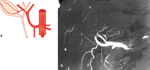
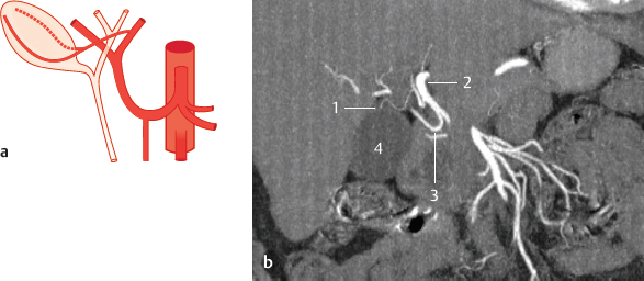
Fig. 15.1 Cystic artery as a branch of the right hepatic artery (46%). Schematic (a) and coronal MIP CT (b). 1 Cystic artery; 2 left hepatic artery; 3 right hepatic artery; 4 gallbladder.
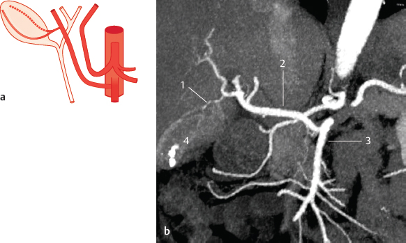
Fig. 15.2 Cystic artery as a branch of an accessory right hepatic artery which originates from the superior mesenteric artery (12%). Schematic (a) and coronal oblique MIP CT (b) (patient with gallstones). 1 Cystic artery; 2 right hepatic artery; 3 superior mesenteric artery; 4 gallbladder with gallstones.
Fig. 15.3 Cystic artery as a branch of the left hepatic artery (5%). Schematic (a) and coronal MIP CT (b). 1 Cystic artery; 2 left hepatic artery; 3 right hepatic artery 4 gallbladder.
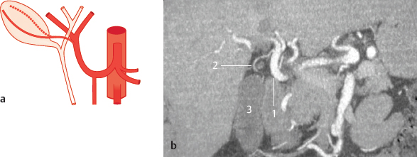
Fig. 15.4 Cystic artery originates from the bifurcation of the common hepatic artery (10%). Schematic (a) and coronal MIP CT (b). 1 Common hepatic artery; 2 cystic artery; 3 gallbladder.
Stay updated, free articles. Join our Telegram channel

Full access? Get Clinical Tree


