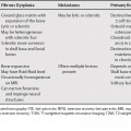
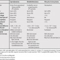
71 A broad range of liver lesions can appear cystic. Hepatocellular carcinoma and hemangioma are the primary liver lesions which are most likely to appear as cystic lesions. Fluid-fluid levels in a lesion are nonspecific and seen in both benign and malignant lesions.1 They are usually secondary to hemorrhage. Ciliated hepatic foregut cysts are histologically similar to mediastinal foregut cysts. They are usually solitary and are most commonly seen in a subcapsular location in the medial segment of the left lobe. The contents of the cysts are variable; the cysts can contain serous fluid, lipid, and mucoid material. On magnetic resonance imaging (MRI), they are hyperintense on T2-weighted magnetic resonance imaging (T2WI), but can have a variable signal intensity on T1-weighted magnetic resonance imaging (T1WI). On computed tomography (CT), they can have high attenuation numbers. The appearance can mimic a solid lesion, but they can be differentiated by their lack of enhancement and characteristic location.2
Cystic Liver Lesions
Less Common Etiologies of Hepatic Cysts
Ciliated Hepatic Foregut Cyst
Undifferentiated Embryonal Cell Sarcoma
Stay updated, free articles. Join our Telegram channel

Full access? Get Clinical Tree


