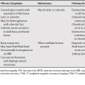
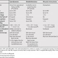
86 Mimics of pancreatic cystic masses, such as duodenal diverticula, should first be excluded. The most important clinical question is whether there has been prior pancreatitis. With a history of pancreatitis, pseudocyst is the most likely diagnosis in most cases. If communication with the pancreatic duct can be demonstrated, or if the duct is dilated, the differential is pseudocyst/chronic pancreatitis and intraductal papillary mucinous tumor (IPMT). Mucinous cystic neoplasm and serous cystadenoma rarely communicate with the duct. In the majority of cases, the main pancreatic duct will be dilated in an IPMT. A main duct IPMT will cause pancreatic ductal dilatation and may mimic the appearance of chronic pancreatitis. A combined (main and side-branch) IPMT can result in both a cystic mass and pancreatic ductal dilatation and can mimic the appearance of chronic pancreatitis and pseudocyst. A side-branch IPMT often is similar in appearance to a combined IPMT because there is often dilatation of the main pancreatic duct due to impaired drainage. If a side-branch IPMT presents without main duct dilatation, the differential diagnosis also might include serous cystadenoma and mucinous cystic neoplasm (however, these entities usually do not communicate with the duct). A history of prior pancreatitis is the primary differentiating factor between a pseudocyst/ chronic pancreatitis and IPMT. IPMT often has internal nodularity, which is not seen in pseudocysts. The appearance of the pancreatic duct can also be helpful in differential diagnosis (Table 86.1). If there is no communication with the duct or duct dilatation, then serous cystadenoma and mucinous cystic neoplasm are the primary considerations (Table 86.2).
Cystic Pancreatic Lesions
Differential Diagnosis of Pancreatic Cystic Lesions
Types of Cystic Pancreatic Masses
Rare Cystic Pancreatic Masses
Pancreatic Cyst Mimics
History of Pancreatitis
Duct Communication/Dilatation
No Duct Communication/Dilatation
| IPMT | Chronic Pancreatitis | |
|---|---|---|
| Duct Strictures | – | + |
| Duct Nodularity* | + | – |
| Papillary Bulging† | + | – |
| Stability | – | + |
| Branch Dilatation | Uncinate, tail | + |
*Secondary to papillary tumor, mucin globs
†More common in malignant IPMT
Imaging Features of Specific Cystic Pancreatic Neoplasms
Mucinous Tumors
Stay updated, free articles. Join our Telegram channel

Full access? Get Clinical Tree


