, Marilyn J. Siegel2, Tomasz Miszalski-Jamka3, 4 and Robert Pelberg1
(1)
The Christ Hospital Heart and Vascular Center of Greater Cincinnati, The Lindner Center for Research and Education, Cincinnati, OH, USA
(2)
Mallinckrodt Institute of Radiology, Washington University School of Medicine, St. Louis, Missouri, USA
(3)
Department of Clinical Radiology and Imaging Diagnostics, 4th Military Hospital, Wrocław, Poland
(4)
Center for Diagnosis Prevention and Telemedicine, John Paul II Hospital, Kraków, Poland
Abstract
The Damus–Kaye–Stansel (DKS) operation is a palliative procedure for patients with a single functioning ventricle with an obstructed rudimentary outflow chamber. It is not used for treatment of hypoplastic left heart syndrome which is treated using the Norwood procedure. operation is used for in double-inlet left ventricle, tricuspid atresia with transposition of the great arteries (TGA), and a common atrioventricular canal with small left ventricular cavity.
The Damus–Kaye–Stansel (DKS) operation is a palliative procedure for patients with a single functioning ventricle with an obstructed rudimentary outflow chamber. It is not used for treatment of hypoplastic left heart syndrome which is treated using the Norwood procedure. This operation is used for in double-inlet left ventricle, tricuspid atresia with transposition of the great arteries (TGA), and a common atrioventricular canal with small left ventricular cavity.
In the DKS operation, the proximal pulmonary artery is divided near its bifurcation and anastomosed to the side of the ascending aorta, thus bypassing any systemic outflow obstruction. Blood flow to the distal pulmonary arteries is reestablished by a modified Blalock–Taussig (preferred in neonates), bidirectional Glenn shunt, or an extracardiac cavopulmonary Fontan procedure (preferred in adults) [1, 2]. Later modifications of the DKS procedure include surgical closure of the aortic valve to prevent development of aortic valve insufficiency [2, 3] and patch augmentation of the aortic arch if the arch is hypoplastic.
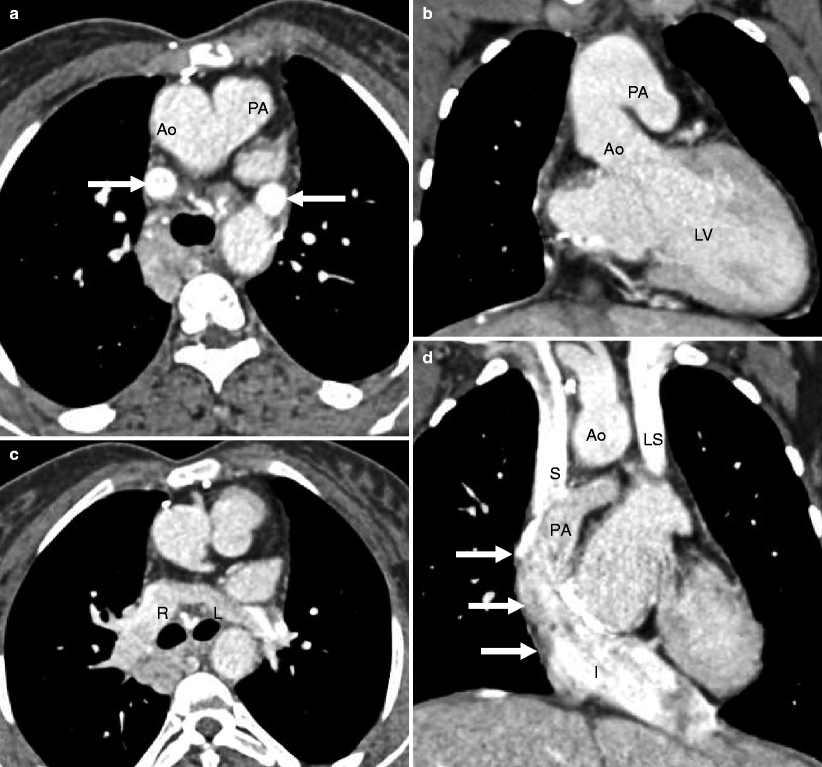
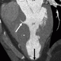
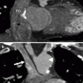
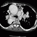
Figures 27.1, 27.2, 27.3, and 27.4 demonstrate the DKS procedure.
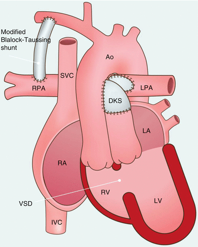

Fig. 27.1
Damus–Kaye–Stansel operation. This diagram shows a surgical connection between the ascending aorta (Ao) and pulmonary artery (PA). The pulmonary artery is ligated just below the anastomotic site. The right and left pulmonary arteries attach to an extracardiac Fontan (FC) conduit (preferred connection in adults, not shown here). The Blalock–Taussig shunt (arrows) is also shown and preferred in infants. The aorta arises from a rudimentary right ventricle. RPA right pulmonary artery, SVC superior vena cava, DKS damus-kaye-stansel anastomosis, LPA left pulmonary artery, LA left atrium, RA right atrium, RV right ventricle, LV left ventricle, VSD ventricular septal defect, IVC inferior vena cava

