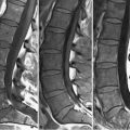
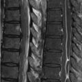
32 Degenerative Disease Longstanding disk herniations or bulges may result in the formation of hypertrophic end plate spurs (i.e., osteophytes). The severity of symptoms resulting from osteophytes (which are typically asymptomatic) does not correlate with their imaging appearance. Osteophyte distribution within the cervical spine directly varies with spinal axis mobility: the mobile lower cervical spine is affected initially with superior spread as disease worsens. Figure 32.1A demonstrates a case of moderately advanced degenerative disease with disk osteophyte complexes at the C3–C7 levels. GRE T2WI aid in distinguishing osteophytes from similarly appearing disk herniations on FSE T2WI: the nucleus pulposus and inner disk annulus, due to their mucopolysaccharide matrix, demonstrate high SI on GRE T2WI, whereas susceptibility effects from calcium in osteophytes result in a very low SI appearance. The osteophytes in the FSE T2WI of Fig. 32.1A result in a mild degree of central canal stenosis—note the lack of CSF surrounding the cord at the involved levels. More severe stenosis is present on the FSE T2WI of Fig. 32.1B where an osteophyte compresses the thecal sac and flattens the cord at the C4–C5 level. The normal AP diameter of the cervical canal (best measured on axial FSE T2WI) is over 13 mm; a canal less than 10 mm in diameter is stenotic. Additional degenerative findings in Fig. 32.1B
![]()
Stay updated, free articles. Join our Telegram channel

Full access? Get Clinical Tree


