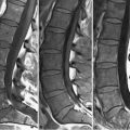
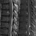
47 Degenerative Disk Disease II Spondylolisthesis is but one cause of central canal stenosis. The sagittal FSE T2WI of Fig. 47.1A illustrates a normally hydrated intervertebral disk at L5–S1, with desiccated disks at all other levels. Despite this, no significant disk bulges or protrusions are present, although the canal is, nevertheless, of small (<11.5 mm) AP diameter. Congenitally shortened pedicles—the etiology of this finding—is evident in the axial T2WI of Fig. 47.1C. A congenitally narrow canal will amplify the severity of any degenerative findings that might develop. The canal in Figs. 47.1B,D is also narrowed. Here, a (B) FSE T2WI demonstrates loss of disk SI at all visualized levels except S1–S2. Loss of vertebral body height at L5–S1 suggests loss of discal SI secondary to degenerative changes rather than normal aging. A prominent disk bulge is also present at L4–L5, which together with ligamentum flavum hypertrophy at this level, results in moderate to severe central canal stenosis—a finding best appreciated on the axial T2WI of Fig. 47.1D. Other structures surrounding the spinal canal can similarly lead to canal stenosis. Degenerative changes of the spine are present in Fig. 47.2
![]()
Stay updated, free articles. Join our Telegram channel

Full access? Get Clinical Tree


