than African Americans. Obesity is associated with a higher incidence of osteoarthritis in the knees, which may be related to an excessive weight-bearing load on these joints.
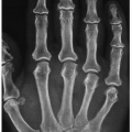
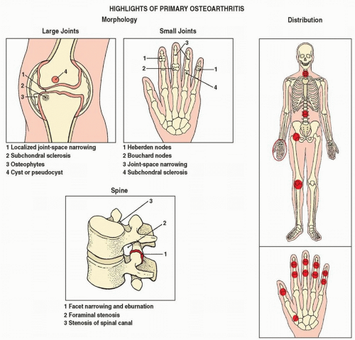 Figure 5.1 ▪ Highlights of the morphology and distribution of arthritic lesions in primary osteoarthritis. |
Table 5.1 CLINICAL AND IMAGING HALLMARKS OF DEGENERATIVE JOINT DISEASE | ||||||||||||||||||||||||||||||||||||||||||||||||||||||||||||||||||||||||||||||||||||||||||||||||||||||||||||||||||||||||||||||||||||||||||||||||||||
|---|---|---|---|---|---|---|---|---|---|---|---|---|---|---|---|---|---|---|---|---|---|---|---|---|---|---|---|---|---|---|---|---|---|---|---|---|---|---|---|---|---|---|---|---|---|---|---|---|---|---|---|---|---|---|---|---|---|---|---|---|---|---|---|---|---|---|---|---|---|---|---|---|---|---|---|---|---|---|---|---|---|---|---|---|---|---|---|---|---|---|---|---|---|---|---|---|---|---|---|---|---|---|---|---|---|---|---|---|---|---|---|---|---|---|---|---|---|---|---|---|---|---|---|---|---|---|---|---|---|---|---|---|---|---|---|---|---|---|---|---|---|---|---|---|---|---|---|---|
| ||||||||||||||||||||||||||||||||||||||||||||||||||||||||||||||||||||||||||||||||||||||||||||||||||||||||||||||||||||||||||||||||||||||||||||||||||||
 Figure 5.2 ▪ Pathology of osteoarthritis of hip joint. Coronal section of the resected femoral head (A) and radiograph of the gross specimen (B) show a large flat osteophyte extending from the medial aspect to the region of fovea (arrows). (Modified from Bullough PG. Atlas of Orthopedic Pathology with Clinical and Radiologic Correlation. 2nd ed. New York, NY: Gower Medical Publishing; 1992, Figs. 10.11 and 10.12, p. 10.5.) |
 Figure 5.3 ▪ Pathology of osteoarthritis of hip joint. Coronal section of the osteoarthritic femoral head shows subchondral bone partially denuded of articular cartilage (arrow). Some articular cartilage remains intact (arrowhead). Observe in the exposed subchondral bone focal bone marrow necrosis (yellow area) because of localized overloading (curved arrow). (Modified from Bullough PG. Atlas of Orthopedic Pathology with Clinical and Radiologic Correlation. 2nd ed. New York, NY: Gower Medical Publishing; 1992, Fig. 9.31.) |
Narrowing of the joint space as a result of thinning of the articular cartilage.
Subchondral sclerosis (eburnation) caused by reparative processes (remodeling).
Osteophyte formation (osteophytosis) as a result of reparative processes in sites not subjected to stress (so-called low-stress areas), which are usually marginal (peripheral) in distribution.
Cyst or pseudocyst formation resulting from bone contusions that lead to microfractures and intrusion of synovial fluid into the altered spongy bone; in the acetabulum, these subchondral cystlike lesions are referred to as Eggers cysts.
 Figure 5.4 ▪ Histopathology of osteoarthritis of hip joint. Photomicrograph of a portion of the articular cartilage of a femoral head shows a fibrous pannus extending over the articular surface (H&E, original magnification ×4). (From Bullough PG. Atlas of Orthopedic Pathology with Clinical and Radiologic Correlation. 2nd ed. New York, NY: Gower Medical Publishing; 1992, Fig. 9.43, p. 9.17.) |
slowly progressive osteoarthritis of the hip is converted into the rapidly progressive, aggressively destructive disease that completely destroys the hip joint. In some of the patients, the major portion of the femoral head may completely disappear. The acetabulum became concentrically enlarged. The pain in the hip is typically disabling and unrelenting. This destructive arthrosis of the hip joint is known as Postel coxarthropathy, a condition characterized by rapid chondrolysis that may quickly lead to complete destruction of the hip joint. Originally described by Lequesne, and also by Postel and Kerboull in 1970, this unique hip disorder occurs predominantly in women, with age of onset at 60 to 70 years. In all cases, a rapid clinical course of hip pain is the consistent common symptom. The histologic findings are those of conventional osteoarthritis with severe degenerative changes in the articular cartilage. However, osteophyte formation is absent or minimal. Hypervascularity in the subchondral bone is a common finding. The bone trabeculae are either abnormally thickened or abnormally thinned. Occasionally, one can observe foci of fibrosis, interstitial edema and hemorrhage in the marrow spaces, focal marrow fat fibrosis, and focal areas of bone resorption. The precise pathogenesis of this condition remains unclear, although direct drug toxicity and the analgesic effects of nonsteroidal anti-inflammatory drugs have been implicated. Some investigators have suggested that intra-articular deposition of hydroxyapatite crystals might lead to joint destruction. Others have proposed subchondral bone ischemia, cell necrosis, and insufficiency fracture of the femoral head as a cause of this arthritis. Some investigators demonstrated elevated levels of interleukin-6 (IL-6) and interleukin-1 beta (IL-1β) in the joint fluid as well as elevated secretion of matrix metal-loproteinases by fibroblasts from the synovium and subchondral cysts. Because of the rapidity of the process, the radiographic presentation of this condition is marked by very little, if any, reparative changes, mimicking infectious or neuropathic arthritis (Charcot joint) (Figs. 5.12, 5.13, 5.14). More recently, Boutry and colleagues reported magnetic resonance imaging (MRI) findings of this form of osteoarthritis. These included joint effusion, a bone marrow edema-like pattern in the femoral head, neck, and acetabulum; femoral head flattening; and cystlike subchondral defects (Fig. 5.15).
 Figure 5.5 ▪ Osteoarthritis of the hip joint. A: A 51-year-old woman presented with a history of right hip pain for the past 10 years and no previous history suggesting predisposing factors for osteoarthritis. Anteroposterior radiograph of the hip demonstrates the radiographic hallmarks of osteoarthritis: narrowing of the joint space, particularly at the weight-bearing segment (arrow), formation of marginal osteophytes (open arrows), and subchondral sclerosis. Note the lack of osteoporosis. B: In another patient, a 65-year-old woman, observe in addition to joint space narrowing, subchondral sclerosis, and osteophytosis, formation of a typical Egger cyst in the acetabulum (arrow). C: Anteroposterior radiograph of the left hip of a 76-year-old man shows narrowing of the joint space mainly at the weigh-bearing segment, subchondral sclerosis, and geode within the femoral head (arrow). D: Conventional radiograph of the right hip joint of a 63-year-old woman with a long history of osteoarthritis demonstrates the classic features of this condition: joint space narrowing, subchondral sclerosis, and osteophytosis. E: Anteroposterior radiograph shows advanced osteoarthritis of both hip joints in this 70-year-old man. |
 Figure 5.6 ▪ CT of osteoarthritis of the hip joint. A: Coronal reformatted image shows diminution of the joint space, osteophytes, and subchondral cysts in the femoral head. B: In another patient, a 69-year-old man, 3D-reconstructed CT image of the pelvis shows advanced osteoarthritis of the right hip joint and moderate OA of the left hip joint. |
 Figure 5.7 ▪ CT of osteoarthritis of the hip joint. A: Anteroposterior radiograph of the left hip of a 66-year-old woman shows narrowing of the joint space, subchondral sclerosis, and cystic-like lesion in the acetabulum, better demonstrated on the coronal reformatted CT image (B). In another patient, a 71-year-old woman, coronal reformatted CT image of the right hip joint (C) shows geodes in the femoral head and acetabulum. D: Coronal reformatted CT image of the left hip joint of a 55-year-old woman shows narrowing of the joint space, subchondral sclerosis, and osteophytosis. |
 Figure 5.8 ▪ CT and MRI of osteoarthritis of the hip joint. A 57-year-old man presented with history of pain in the right groin and “locking” and “clicking” in the hip joint. A: Conventional radiograph shows advanced osteoarthritis of the hip joint. There is suggestion of an osteochondral body in the medial joint compartment, better demonstrated on the coronal reformatted CT image (B). C: Coronal T2-weighted fat-suppressed MR image shows in addition a large joint effusion. |
 Figure 5.9 ▪ MRI of osteoarthritis of the hip. Coronal proton density-weighted fat-suppressed MR image of the right hip of a 68-year-old woman shows joint space narrowing, subchondral sclerosis and bone marrow edema, osteophytes arising from the femoral head and acetabulum, and joint effusion. |
 Figure 5.10 ▪ Migration of the femoral head. A: Superolateral migration of the femoral head is present in this 59-year-old woman with advanced osteoarthritis of the right hip joint. B: Another example of superolateral migration of the femoral head in an 80-year-old man. C: Medial migration of the femoral head is apparent in this 48-year-old woman with osteoarthritis of the right hip. D: Another example of medial migration of the femoral head in a 70-year-old man. E: Axial migration of the femoral head is evident in this 57-year-old woman who was suspected of having inflammatory arthritis. Clinical and laboratory investigations, however, led to a diagnosis of idiopathic osteoarthritis, which was confirmed on histopathologic examination after total hip replacement. |
 Figure 5.11 ▪ Rheumatoid arthritis with superimposed secondary osteoarthritis. Anteroposterior radiograph of the right hip of a 42-year-old woman with a known history of long-standing rheumatoid arthritis shows the typical changes of inflammatory arthritis, including axial migration of the femoral head and acetabular protrusio. Superimposition of secondary osteoarthritis is evident in subchondral sclerosis and formation of marginal osteophytes. |
joint, particularly flexion and internal rotation; and (c) imaging findings on conventional radiography, CT, and MRI. In cam type, conventional radiography demonstrates excessive bone formation at the femoral head/neck junction with loss of normal anatomic “waist” at this site (Fig. 5.25A), occasionally resembling the smooth hand grip of some pistols (“pistol grip deformity” or a “cam effect”) (Fig. 5.25B); an os acetabulum, which more likely represents an osseous metaplasia of the cartilaginous labrum or a fragment of damaged acetabular rim; and a radiolucent lesion at the head/neck junction, formerly called synovial herniation pit, and now designated as fibroosseous lesion. CT shows these abnormalities even better (Fig. 5.26). MR arthrography (MRa), particularly the radial reformatted images, in addition to the findings listed previously, clearly demonstrates abnormalities of the fibrocartilaginous labrum at the anterosuperior portion of the acetabulum (Fig. 5.27; see also Fig. 2.91). In pincer type, particularly in case of acetabular retroversion, conventional radiograph shows “crossover” sign, when more lateral projection of anterior acetabulum, which normally should project medially to the posterior acetabulum, “crosses” the
posterior acetabular outline (Fig. 5.28). MRI demonstrates acetabular version and depth of the femoral head coverage (Fig. 5.29). To determine the sphericity of the femoral head and the prominence of the anterior femoral head/neck junction, the alpha angle is calculated on the oblique axial CT or oblique axial MR images (Fig. 5.30). Radial reformatted MR images are of particular value in this respect because they allow optimal visualization of the anterosuperior region of the femoral head/neck junction, where the most significant changes in the alpha angle occur (see Fig. 5.27). The normal alpha angle should not exceed 50 degrees. The larger the alpha angle, the more pronounced is nonspherical shape of the femoral head, and the greater is predisposition for anterior FAI.
 Figure 5.12 ▪ Postel coxarthropathy. A: Anteroposterior radiograph of the right hip of a 72-year-old man who had pain in the hip for 4 months shows the typical appearance of this arthrosis, which often mimics Charcot joint or infectious arthritis. Note the destruction of the articular portion of the femoral head, which is flattened and laterally subluxed. There is eccentric depression of the lateral articular surface. The same destructive process has led to widening of the acetabulum. B: Similar deformity of the right hip joint is seen in this 69-year-old woman. Note relative absence of osteophytes. |
 Figure 5.13 ▪ Postel coxarthropathy. A: Osteoarthritis of the right hip joint in this 61-year-old woman markedly progressed in a very short time as seen on the radiograph obtained 5 months later (B). |
 Figure 5.14 ▪ CT of Postel coxarthropathy. Coronal reformatted CT image of the right hip shows characteristic features of this destructive arthritis: marked deformity of the femoral head with flattening of the subchondral segment assuming hatchet-like appearance, widening of the acetabulum, and relatively only mild reactive changes. |
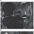
 Figure 5.15 ▪ Arthrography and MRI of Postel coxarthropathy. A: Anteroposterior radiograph of the right hip of a 44-year-old man shows destructive changes of the femoral head and acetabulum. B: Aspiration arthrogram, which was performed to rule out infection, shows hypertrophic synovitis. C: Gradient echo T2*-weighted MRI shows joint effusion, hypertrophied synovium, and subchondral cysts in the acetabulum and femoral head. |
 Figure 5.16 ▪ Posttraumatic osteoarthritis. Anteroposterior radiograph of the pelvis of a 40-year-old man, who 7 years prior to this examination sustained a complex fracture of the right proximal femur and acetabulum, shows a deformity of the femoral head and neck associated with narrowing of the hip joint space, subchondral sclerosis, and osteophyte formation. Note the completely normal left hip joint. |
 Figure 5.17 ▪ CT of posttraumatic osteoarthritis. A 64-year-old man, who in the past sustained complex right acetabular and femoral fractures, developed secondary osteoarthritis. A: Preliminary scout CT image shows posttraumatic deformity of the acetabulum and femoral head associated with acetabular protrusio. B: Axial CT section through both hips shows osteoarthritic changes of the right femoral head and ununited fracture of the anterior column (arrow). C: Coronal reformatted image demonstrates significant narrowing of the joint space, deformity of the femoral head, and periarticular sclerosis. All CT features are consistent with posttraumatic osteoarthritis. |
 Figure 5.18 ▪ Secondary osteoarthritis because of slipped capital femoral epiphysis (SCFE). A: Anteroposterior radiograph of the pelvis of a 40-year-old man with history of SCFE shows osteoarthritic changes of the left hip joint. Note osseous remodeling at the junction of the femoral head and neck, a hallmark of this condition, known as a Herndon hump (arrow). B: In another patient, a 24-year-old woman with OA of the left hip joint, observe a characteristic Herndon hump (arrow) pointing to SCFE as the underlying cause of arthritis. |
 Figure 5.19 ▪ Secondary osteoarthritis because of developmental dysplasia of the hips (DDH). Coronal reformatted (A) and 3D reconstructed with surface-rendering algorithm (B) CT images of the hips of a 30-year-old woman with history of bilateral congenital hip dislocation show dysplastic changes of both hip joints. There is evidence of secondary osteoarthritis manifested by narrowing of the joint spaces, subchondral sclerosis at the site of femoral heads and acetabula, and formation of small marginal osteophytes at the periphery of both acetabula. |
 Figure 5.20 ▪ Secondary osteoarthritis because of osteonecrosis. A 48-year-old man, a chronic alcoholic, developed osteonecrosis of both femoral heads, marked by increased bone density and subchondral collapse. Secondary osteoarthritis is distinguished by narrowing of the joint space, marginal osteophytosis, and subchondral cyst formation. |
 Figure 5.21 ▪ Secondary osteoarthritis because of Paget disease. A: Radiograph of the pelvis of an 80-year-old woman shows cool phase of Paget disease affecting pelvic bones and both femora. Note advanced osteoarthritis of both hip joints with almost complete obliteration of the joint spaces. B: In another patient, a 75-year-old woman with long-standing polyostotic Paget disease, anteroposterior radiograph of the right hip demonstrates advanced osteoarthritis associated with acetabular protrusio. |
 Figure 5.22 ▪ Secondary osteoarthritis because of joint infection. Anteroposterior radiograph of the pelvis of a 49-year-old man with history of septic arthritis of the right hip joint and acetabular osteomyelitis shows deformity of the acetabulum, subchondral sclerosis, and significant narrowing of the joint space. Note unremarkable left hip joint. |
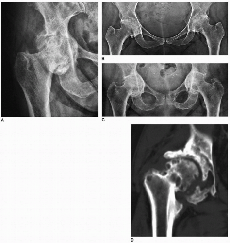 Figure 5.23 ▪ Secondary osteoarthritis because of inflammatory arthritis (RA). A: Radiograph of the right hip of a 60-year-old woman shows concentric narrowing of the joint space and acetabular protrusio, features of inflammatory arthritis. Superimposed are features of osteoarthritis comprising sclerotic changes of the femoral head and acetabulum and osteophytosis. B: In another patient, a 38-year-old woman with bilateral hip rheumatoid arthritis, observe typical features of inflammatory arthritis and secondary osteoarthritic changes manifesting mainly by formation of prominent osteophytes. C: Similar example of secondary osteoarthritis superimposed on rheumatoid arthritis affecting both hip joints is seen in an 81-year-old woman. D: Coronal reformatted CT image of the right hip of a 40-year-old woman with rheumatoid arthritis shows subchondral and osseous erosions of the femoral head and acetabulum and super-imposed secondary osteoarthritic changes in form of subchondral sclerosis of the acetabulum and osteophyte formation. |
 Figure 5.24 ▪ Secondary osteoarthritis because of inflammatory arthritis (psoriasis). Radiograph of the pelvis of a 64-year-old man with clinically documented psoriasis shows characteristic for inflammatory arthritis concentric narrowing of the hip joints and axial migration of the femoral heads. In addition note the changes of super-imposed secondary osteoarthritis marked by subchondral sclerosis, osteophytosis, and cyst formation in the left acetabulum and in the right femoral head. The patient also developed left-sided sacroiliitis (arrow). The arrowhead points to unaffected right sacroiliac joint. |
 Figure 5.25 ▪ Cam type of FAI. A: Anteroposterior radiograph of the right hip of a 39-year-old woman shows excessive bone buildup at the femoral head/neck junction (arrow). Note secondary osteoarthritis of the hip joint. B: In another patient, a 41-year-old man, tubular appearance of the proximal right femur and the osseous prominence at the femoral head/neck junction assumed a “pistol grip” deformity. Also evident is osteoarthritis of the hip joint. |
 Figure 5.26 ▪ CT of cam type FAI. Coronal reformatted (A) and 3D-reconstructed (B) CT image in shaded surface display in a 34-year-old man show bone accretion at the femoral head/neck junction (arrows). |
 Figure 5.27 ▪ MR arthrography of cam type FAI. Radial reconstructed MRa images of the hip joint show various characteristic features of this abnormality. A: In a 34-year-old woman—a decreased femoral head/neck offset associated with hypertrophic ossification (arrow). B: In a 32-year-old woman—a fibroosseous lesion at the anterosuperior aspect of the femoral head/neck junction (arrow). C: In a 38-year-old man—a tear of the superior anterior cartilaginous labrum (arrow). D: In a 30-year-old woman—a delamination injury to the acetabular labrum (arrow). |
gross deformities of the knees are become obvious, such as valgus or varus configuration (Fig. 5.31).
 Figure 5.28 ▪ Pincer type FAI. A: Anteroposterior radiograph of the left hip in a 29-year-old woman shows a crossover sign. Note that the posterior acetabular rim outline (yellow line) projects medially (arrow) in relation to the anterior acetabular rim (red line), indicative of acetabular retroversion. B: In a normal hip joint, the posterior acetabular rim outline projects laterally to the anterior acetabular rim. |
 Figure 5.29 ▪ MRI of pincer FAI. A: Axial oblique T1-weighted MR image shows deeply seated femoral head secondary to acetabular retroversion. Acetabular depth can be quantified by drawing a line (ab) connecting the posterior and anterior acetabular rims and a parallel line (cd) that passes through the center of the femoral head (red dot). The distance between these two lines defines the acetabular depth, with the value being positive (+) if the center of the femoral head projects lateral to the line connecting the acetabular rims. Negative values (-) indicate deep seating of the femoral head within the acetabulum. B: Axial oblique MR image of normal hip joint is shown for comparison. |
 Figure 5.30 ▪ Femoroacetabular impingement—calculation of alpha angle. The alpha angle is formed by the intersection of two lines: line AB, drawn from the center of the femoral head (A) to the point where peripheral osseous contour of the anterior femoral head intersects the extrapolated circle of the femoral head (B), and the second line AC, drawn from the center of the femoral head (A) through the longitudinal axis of the femoral neck (C). Normal alpha angle should not exceed 50 degrees. A: Alpha angle calculated on the oblique axial CT image of the right hip in a patient with cam FAI. B: Alpha angle calculated on the oblique axial MR image of the left hip in a patient with cam FAI. The arrows point to excessive bone formation at the anterosuperior aspect of the femoral head/neck junction. |
 Figure 5.31 ▪ Knee deformities. Patient with advanced osteoarthritis of the knee joints affecting predominantly the medial compartments developed varus deformities. |
Related posts:


Stay updated, free articles. Join our Telegram channel

Full access? Get Clinical Tree


