Presentation and Presenting Images
( ▶ Fig. 50.1, ▶ Fig. 50.2, ▶ Fig. 50.3, ▶ Fig. 50.4, ▶ Fig. 50.5, ▶ Fig. 50.6)
A 72-year-old female presents for routine screening mammography.
50.2 Key Images
( ▶ Fig. 50.7, ▶ Fig. 50.8, ▶ Fig. 50.9, ▶ Fig. 50.10, ▶ Fig. 50.11, ▶ Fig. 50.12
50.2.1 Breast Tissue Density
There are scattered areas of fibroglandular density.
50.2.2 Imaging Findings
The imaging of the right breast is normal (not shown). The left breast demonstrates an increasing or developing asymmetry at the 1 to 2 o’clock position in the posterior depth, 8 cm from the nipple ( ▶ Fig. 50.7 and ▶ Fig. 50.8 and the mammogram comparisons ▶ Fig. 50.9 and ▶ Fig. 50.10). Tomosynthesis confirms the finding and shows it on the left craniocaudal (CC) tomosynthesis movie slice 26 of 62 ( ▶ Fig. 50.11) and on the left mediolateral oblique (MLO) tomosynthesis movie slice 24 of 69 ( ▶ Fig. 50.12).
50.3 BI-RADS Classification and Action
Category 0: Mammography: Incomplete. Need additional imaging evaluation and/or prior mammograms for comparison.
50.4 Diagnostic Images
( ▶ Fig. 50.13)
50.4.1 Imaging Findings
The ultrasound demonstrated a small group of cysts (circle) within a focal asymmetry of fibroglandular tissue, 6.75 cm from the nipple (dashed line in ▶ Fig. 50.13). This correlates with the mammographic finding.
50.5 BI-RADS Classification and Action
Category 2: Benign
50.6 Differential Diagnosis
Cysts: A developing asymmetry has an increased risk for malignancy but may be a benign finding, as in this case. In this situation, this small group of cysts developed in this focal asymmetry of fibroglandular tissue causing this to appear more prominent.
Hematoma: A developing asymmetry that is due to a hematoma needs to have an appropriate clinical history of trauma or a medical procedure proceeding the development of the finding. This patient had no history of trauma.
Breast cancer: Developing asymmetries are not common, however are associated with an increased likelihood of malignancy. Imaging should be performed to ensure if the developing asymmetry is benign or malignant. In this case, the ultrasound confirms that the finding is benign.
50.7 Essential Facts
On mammography, a developing asymmetry is defined as a focal asymmetry that has appeared or increased in size or conspicuity since a previous examination.
Obtaining a conventional two-dimensional (2D) mammogram and tomosynthesis at the same time allowed direct comparison between the 2D mammogram and the tomosynthesis images. The comparison helped confirm that the finding is a developing asymmetry.
Developing asymmetries are uncommon. When this finding is identified on screening and diagnostic mammography, the likelihood of malignancy is sufficiently high to justify recall and biopsy. The likelihood of malignancy is 15% at screening mammography and 25% at diagnostic mammography.
Sonography is useful to confirm or exclude malignancy and to guide biopsy.
Normal sonographic findings do not exclude malignancy in the case of a developing asymmetry.
50.8 Management and Digital Breast Tomosynthesis Principles
Tomosynthesis helped to identify and confirm this finding because of its ability to remove the effects of overlapping, obscuring breast tissue.
Tomosynthesis’s ability to unmask lesions increases the number of masses seen on mammography. Not all of these unmasked lesions will be cancer. The morphology of each lesion needs to be assessed.
With DBT, lesions are unmasked irrespective of the density of the breast tissue.
When no suspicious findings are seen on ultrasound, tomosynthesis-directed stereotactic biopsy can be performed.
50.9 Further Reading
[1] Leung JW, Sickles EA. Developing asymmetry identified on mammography: correlation with imaging outcome and pathologic findings. AJR Am J Roentgenol. 2007; 188(3): 667‐675 PubMed
[2] Sickles EA, D’Orsi CJ, Bassett LW, et al. ACR BI-RADS Mammography. In: ACR BI-RADS Atlas, 5th edition. Reston, VA: American College of Radiology; 2013
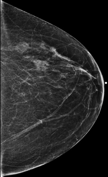
Fig. 50.1 Left craniocaudal (LCC) mammogram.
Related posts:


Stay updated, free articles. Join our Telegram channel

Full access? Get Clinical Tree


