1. Benign epithelial tumors
Pleomorphic adenoma
Myoepithelioma
Basal cell adenoma
Warthin tumor
Oncocytoma
Canalicular adenoma
Sebaceous adenoma
Lymphadenomas: sebaceous and nonsebaceous
Ductal papilloma
Cystadenoma
2. Malignant epithelial tumors
Acinic cell carcinoma
Mucoepidermoid carcinoma
Adenoid cystic carcinoma
Polymorphous low-grade adenocarcinoma
Epithelial-myoepithelial carcinoma
Clear cell carcinoma, not otherwise specified
Basal cell adenocarcinoma
Sebaceous carcinoma
Sebaceous lymphadenocarcinoma
Cystadenocarcinoma
Mucinous adenocarcinoma
Oncocytic carcinoma
Salivary duct carcinoma
Adenocarcinoma, not otherwise specified
Myoepithelial carcinoma
Carcinoma ex pleomorphic adenoma
Carcinosarcoma
Metastasizing pleomorphic adenoma
Squamous cell carcinoma
Small cell carcinoma
Large cell carcinoma
Lymphoepithelial carcinoma
Sialoblastoma
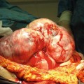
Amongst benign parotid gland tumors, polymorphous and Warthin’s tumors account for 90 % of cases. Benign histopathological Type III is a basal cell gland tumor, accounting for 3–4 % of cases. Malignant parotid gland tumors have the following characteristics. (1) Histopathological types vary, and have characteristic tumor activity. Moreover, even for the same histopathological type, the degree of malignancy varies from low to high. (2) There are many low-malignancy cases. Therefore, long-term observation is important. (3) Preoperative histopathological diagnosis is difficult, and the only method for establishing a preoperative diagnosis is Fine-Needle Aspiration cytology (FNA). However, the accuracy of this type of diagnosis is low.
It was reported that the proportion of accessory parotid gland cases of all parotid gland tumors in the 1960s was 7.7 % [3]; however, a subsequent report only states 1 % [4], therefore it is believed that accessory parotid gland tumors are relatively rare. The same type of tumor may develop either in the accessory parotid gland or parotid gland; however, while the incidence rate of malignant parotid gland tumor is 25 %, that of malignant accessory parotid gland tumor is 42–55 %, which is higher [4, 5]. Many benign tumors are polymorphous, and many malignant tumors are mucoepidermoid. Studies state that the proportion of mucoepidermoid tumors is higher in accessory parotid gland tumors than in parotid gland tumors. It is assumed that one of the reasons for this is that accessory parotid glands have many mucous glands.
27.3 Diagnosis of Accessory Parotid Gland Tumors
It is important to suspect the possibility of an accessory parotid gland tumor firstly based on its anatomical position. A necessary condition for a tumor to be an accessory parotid gland tumor is for imaging examinations to show no continuity with the parotid gland tissue on CT and MRI. A confident diagnosis can be made if secretory ducts flowing into Stensen’s duct can be observed on images such as sialography, sialo-CT and MR- sialography. However, even if secretory ducts can not be observed, the possibility of a tumor derived from the accessory parotid gland cannot be completely ruled out. FNA is useful in observing salivary gland tissue and in qualitative diagnosis [6]; however, it is necessary to be aware that there are cases of false positives and false negatives. A definitive diagnosis should be made based on an isolated sample.
27.4 Differential Diagnosis
Diseases which should be differentially diagnosed from accessory parotid gland tumors are tumors developing in the cheek region, including benign tumors such as schwannoma, dermoid cyst, and lipoma; as well as lymph node and masseter muscle tumors, Stensen’s duct primary tumors, minor salivary gland derived tumors, and aberrant salivary gland tumors. The histological subtypes of these tumors can be differentiated based on histopathological findings; however, tissues from which the latter four types of tumors are derived can be identified by employing the following examinations.
1.
Parotid gland and accessory parotid gland tumors can be differentiated based on whether continuity can be observed between the tumor and the parotid gland.
2.
Stensen’s duct primary tumors and accessory parotid gland tumors can be differentiated based on whether histopathological invasion of tumor cells into Stensen’s duct can be observed.
3.
If the tumor is positioned on the lateral side of the masseter muscle, the possibility of it being a minor salivary gland primary tumor is extremely low.