Figure 1.1 ▪ Classification of the arthritides and arthropathies. CPPD, calcium pyrophosphate deposition; MCTD, mixed connective tissue disease. |
may point to the diagnosis of interphalangeal osteoarthritis; swelling of the proximal and distal interphalangeal joints (Fig. 1.4A) and nail changes such as onycholysis (separation of the nail plate from the nail bed) (Fig. 1.4B), pitting of the nails (Fig. 1.4C), leukonychia (white nails, milk spots) (Fig. 1.4D), splinting hemorrhages (Fig. 1.4E), subungual hyperkeratosis (Fig. 1.4F), and swelling of the entire finger (dactylitis or “sausage digit”) (Fig. 1.4G) are commonly seen in psoriasis; flexible, easy-reducible joint contractures combined with redness and telangiectasia of the nail fold capillaries may be indicative of systemic lupus erythematosus; tapering of the ends of digits and sclerodactyly is characteristic of scleroderma. In the foot, the redness and swelling of the big toe may indicate the presence of gout (Fig. 1.5).
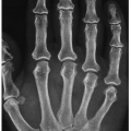
 Figure 1.2 ▪ Rheumatoid arthritis. Observe ulnar deviation in the swollen metacarpophalangeal joints of both hands and hyperextension of the thumbs (“hitchhiker’s thumbs”). |
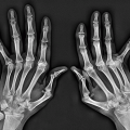
 Figure 1.3 ▪ Rheumatoid arthritis. A: Flexion in the proximal interphalangeal joints and extension in the distal interphalangeal joints of the index and middle fingers resulted in boutonnière deformity. B: In another patient, note characteristic boutonnière deformity of the index finger. C: Extension in the proximal and flexion in the distal interphalangeal joint is known as a swan-neck deformity. |
 Figure 1.4 ▪ Psoriatic arthritis. A: Note swelling of the proximal and distal interphalangeal joints and skin changes on the index finger. B: Detachment of the nail from the underlying nail bed is termed onycholysis. C: The surface of the nail develops small pits that look like a surface of a thimble (nail pitting). The number of the pits can vary from a few to dozens. D: White spots on the nail are termed leukonychia. E: Reddish-brown lines and spots, termed splinter hemorrhage, represent tiny blood clots that run vertically under the nail because of damage of small capillaries. F: Typical appearance of hyperkeratosis, a chalky substance that accumulates under the nail. G: Diffusely swollen thumb (sausage digit) represents a dactylitis. Note also typical for psoriasis skin changes. |
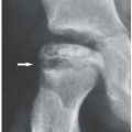
 Figure 1.5 ▪ Gouty arthritis. Observe the big toe swelling and dorsal mass at the site of the first metatarsophalangeal joint representing a tophus. (Reprinted from Klein MJ, Bonar SF, Freemont T, et al., eds. Atlas of Nontumor Pathology. Non-neoplastic Diseases of Bones and Joints. Washington, DC: American Registry of Pathology and Armed Forces Institute of Pathology; 2011, Fig. 8-206, with permission of the author and the Publishers.) |
 Figure 1.6 ▪ Synovial fluid. Depending upon the content, the synovial fluids exhibit different colors. From left to right: normal, noninflammatory arthropathy, hemarthrosis, xanthochromic, and ochronotic. (Reprinted from Klein MJ, Bonar SF, Freemont T, et al., eds. Atlas of Nontumor Pathology. Non-neoplastic Diseases of Bones and Joints. Washington, DC: American Registry of Pathology and Armed Forces Institute of Pathology; 2011, Fig. 8-27, with permission of the author and the Publishers.) |
effusion is in doubt. In most diagnostic and all therapeutic procedures, an attempt should be made to withdraw all fluid accumulated in the joint space. Nonetheless, although about 5 mL is sufficient for most of the necessary studies, as little as one drop may be diagnostic of gout or pyrophosphate dehydrate crystal deposition disease if the appropriate crystals are present (see Figs. 1.9, 1.10, 1.11, 1.12). Furthermore, most studies can be minimized to scale down the amount of drowned fluid in pediatric patients. If there is doubt regarding the origin of fluid from a tap (i.e., blood vs. bloody tap or subcutaneous fluid vs. synovial fluid), this may be resolved with the mucin test or metachromatic staining for the presence of hyaluronate (Table 1.4).
Table 1.1 CATEGORIES OF JOINT FLUID AND DISEASE ASSOCIATIONS | ||||||||||||||||||||||||||||||||||||||||||||||||||||||||||||||||||
|---|---|---|---|---|---|---|---|---|---|---|---|---|---|---|---|---|---|---|---|---|---|---|---|---|---|---|---|---|---|---|---|---|---|---|---|---|---|---|---|---|---|---|---|---|---|---|---|---|---|---|---|---|---|---|---|---|---|---|---|---|---|---|---|---|---|---|
|
 Figure 1.7 ▪ Synovial fluid—rheumatoid arthritis. A: Clotted fibrin viewed between polarizers in a wet preparation of synovial fluid obtained from a patient with active rheumatoid arthritis. B: Ragocytes viewed in diffused light in a wet preparation of synovial fluid from the same patient. Observe characteristic large granules within the ragocytes, representing cytoplasmatic inclusions of ingested aggregated IgG immunoglobulin and fibrin. (Reprinted from Klein MJ, Bonar SF, Freemont T, et al., eds. Atlas of Nontumor Pathology. Non-neoplastic Diseases of Bones and Joints. Washington, DC: American Registry of Pathology and Armed Forces Institute of Pathology; 2011, Fig. 8-190, with permission of the author and the Publishers.) |
 Figure 1.8 ▪ Synovial fluid—hydroxyapatite crystals. The normally almost invisible crystalloids of hydroxyapatite within a fibrin clot form birefringent red complexes with the alizarin when stained with alizarin red. (Reprinted from Klein MJ, Bonar SF, Freemont T, et al., eds. Atlas of Nontumor Pathology. Non-neoplastic Diseases of Bones and Joints. Washington, DC: American Registry of Pathology and Armed Forces Institute of Pathology; 2011, Fig. 8-34, with permission of the author and the Publishers.) |
 Figure 1.9 ▪ Synovial fluid—pyrophosphate crystals. Pyrophosphate crystals (blue and red) are identified by polarizing microscopy with interference (compensating) plate in the path of light (H&E, polarized light). (Reprinted from Klein MJ, Bonar SF, Freemont T, et al., eds. Atlas of Nontumor Pathology. Non-neoplastic Diseases of Bones and Joints. Washington, DC: American Registry of Pathology and Armed Forces Institute of Pathology; 2011, Fig. 8-16, with permission of the author and the Publishers.) |
 Figure 1.10 ▪ Synovial fluid—pyrophosphate crystals. A: Pyrophosphate crystals within the synovial fluid cytocentrifuge stained with Jenner-Giemsa. The unstained spaces contain the crystals. B: Same specimen viewed now in polarized light with a quarter-wave plate shows the crystals more clearly. (Reprinted from Klein MJ, Bonar SF, Freemont T, et al., eds. Atlas of Nontumor Pathology. Non-neoplastic Diseases of Bones and Joints. Washington, DC: American Registry of Pathology and Armed Forces Institute of Pathology; 2011, Fig. 8-222, with permission of the author and the Publishers.) |
 Figure 1.11 ▪ Synovial fluid—urate crystals. A: The needle-shaped monosodium urate monohydrate crystals in synovial fluid aspirated from a patient with acute gouty arthritis are viewed here in polarized light with a quarter-wave compensating plate. The slow wave vector (the direction of the extraordinary axis of the compensating plate) is indicated by the arrow. B: In the synovial fluid obtained from another patient with acute gouty arthritis and viewed in polarizing light with an interposed quarter-wave plate, the urate crystals are clearly visible. C: Urate crystals within a neutrophil (Jenner-Giemsa stain). (A and C reprinted from Klein MJ, Bonar SF, Freemont T, et al., eds. Atlas of Nontumor Pathology. Non-neoplastic Diseases of Bones and Joints. Washington, DC: American Registry of Pathology and Armed Forces Institute of Pathology; 2011, Figs. 8-31 and 8-213, with permission of the author and the Publishers.)
Related posts:Stay updated, free articles. Join our Telegram channel
Full access? Get Clinical Tree
 Get Clinical Tree app for offline access

|