63 Diffusion-Weighted Imaging
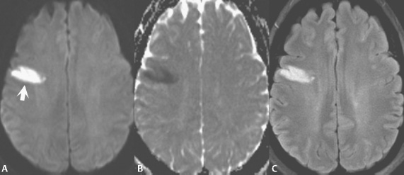
Fig. 63.1
An early subacute, right middle cerebral artery (MCA) infarct is illustrated in Fig. 63.1 (arrow). Images were acquired with diffusion gradients applied in the phase, read, and slice directions and combined to form a single composite diffusion-weighted image (Fig. 63.1A), also known as a trace-weighted image. This image has both diffusion and T2 weighting. A parameter specific to diffusion-weighted imaging (DWI) is the b-value, which defines the strength of the applied diffusion gradient. Increasing the b-value increases the diffusion weighting and thus, up to a point, the conspicuity of early infarcts, but with a concomitant decrease in signal-to-noise ratio. A typical b-value for modern 1.5 and 3 T scanners is 1000 sec/mm2. To obtain an image with only diffusion information, at least one additional scan has to be acquired, specifically using the same technique but without the application of diffusion gradients (b = 0). This scan is then used in conjunction with the diffusion-weighted scan to calculate an apparent diffusion coefficient (ADC) map (Fig. 63.1B), in which there is no T2 weighting. A lesion with restricted diffusion, such as an early infarct (due to the presence of cytotoxic edema), has high signal intensity (SI) on diffusion-weighted images and low SI on the ADC map, the latter reflecting to the low diffusion coefficient. The fluid-attenuated inversion recovery (FLAIR) scan, Fig. 63.1C, in this instance also visualizes the MCA infarct well, but for a different reason, the prolongation of T2 due to the presence of vasogenic edema.
Stay updated, free articles. Join our Telegram channel

Full access? Get Clinical Tree


