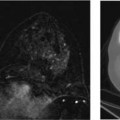
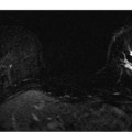
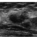
5 Digital Mammographic Appearance of Benign and Malignant Calcifications Detecting breast microcalcifications is one of the most important ways to identify breast cancer. Fifty to 80% of breast cancers have microscopic calcifications.1 Furthermore, clustered microcalcifications consist of up to 40% of non-palpable cancers.2 Because the spatial resolution of digital mammography is poorer than screen-film mammography, there has been concern that digital mammography is less sensitive in displaying malignant microcalcifications compared with screen-film. Whereas the spatial resolution of screen-film mammography is greater than 15 line pairs per mm (lp/mm), digital mammography units have been found to have a spatial resolution as low as 5 lp/mm. Despite the reduced spatial resolution, studies have reported that digital mammographic units display simulated microcalcifications within phantoms in a manner that is at least comparable to screen-film machines.3 This relative equivalence in diagnostic detectability applies to the comparison of digital spot mammography with conventional spot mammography.4 Researchers from the M. D. Anderson Cancer Center (Houston, Texas) have found that within a breast phantom that has a uniform background, digital flat-panel mammographic units perform better than conventional screen-film mammography units in identifying particles that simulate microcalcifications.5 After magnification is added to the imaging devices, the performance of conventional screen-film equipment becomes equivalent to digital flat-panel mammography. When the simulated calcifications are placed within a phantom with a background that simulates complex breast tissue, the screen-film and the digital charge-coupled device unit initially perform better. However, magnification substantially improves the performance of the digital flat-panel mammographic unit, so that there is no significant difference. Although microcalcifications may be visible with larger pixel digital images, Ruschin et al noted that increasing pixel size is associated with decreasing the ability of radiologists to identify the shape of the microcalcifications.6 Multiple retrospective studies have been performed to compare the visibility of microcalcifications using digital mammography with conventional screen-film mammography. Fischer and colleagues compared the appearance of 37 cases of microcalcifications (21 malignant, 16 benign) that had been documented with screen-film mammography and digital mammography hard copy filmed images.7 They reported that all breast imagers had a slightly higher sensitivity and specificity for the calcifications using the digital mammographic images compared with the screen-film images, but this difference was not statistically significant. Multiple clinical investigators using soft copy review of digital images have reinforced these findings: receiver operator characteristic analysis indicates that the performance of breast imagers using digital mammography is better compared with screen-film. However, these differences are not statistically significant.8–10 As radiologists become more experienced and as the equipment advances, digital mammographic evaluation of calcifications will continue to improve. Kim et al reported that after comparing the image quality, number, and conspicuity of 40 clustered calcifications (3 malignant, 37 benign), radiologists judged soft copy digital mammographic images as better in all categories compared with screen-film exams.11 When using digital mammography, a routine method to evaluate the breast for microcalcifications should be incorporated into screening. If hard copy film is being used to review digital mammograms, the routine will probably be similar to the screen-film mammographic method. However, if a soft copy display is being used, then a digital imaging method is preferable. The initial identification may involve a rapid review of the four screening views using a presentation size that is at least equivalent to that used in mammographic film. Because of the flexibility of soft copy display, the screening views can be displayed at a size larger than film. Once the screening views are reviewed, the display can be magnified to specifically identify calcifications. This manipulation would be comparable to using a magnification glass with screen-film. Before enlarging or magnifying the image, the radiologist may prefer to superimpose a proprietary higher contrast display mode or a high-resolution mode. Magnification may be performed by sweeping the image with a small field of view (FOV) digital magnifying glass or by magnifying large segments of the image (e.g., a quadrant or a third of the image). Computer-aided detection (CAD) is commonly used in conjunction with digital mammographic screening exams. Although each CAD system uses different labels, all manufacturers have annotations that denote the presence of possible microcalcifications. Multiple studies have shown that CAD programs tend to perform better in identifying malignant microcalcifications compared with masses.12,13 Retrospective studies have suggested that CAD has an excellent receiver operating characteristics curve and that it may reduce interoperator variability.14–16 More experienced readers probably benefit less from CAD than do less experienced interpreters.17 However, even with the use of CAD, the radiologist still bears the primary responsibility for identifying suspicious calcifications, as CAD is not sensitive in identifying less conspicuous depositions, such as amorphous calcifications.18 CAD results can be viewed either at the beginning or at the end of the screening examination. By viewing the CAD images early, the radiologist can incorporate the examination of any suspected CAD calcifications into a routine screening evaluation. If CAD images are viewed at the end of the screening examination, the radiologist should plan to reevaluate the breast for CAD-identified calcifications that were initially missed. With experience, the radiologist will miss few CAD-identified significant microcalcifications. Once calcifications are identified, digital mammography offers an array of new methods to examine microcalcifications. In general, when examining calcifications, the highest resolution available on a particular system should be used. Furthermore, a display mode should be used that allows the operator to characterize the shape of the calcifications. Alternatively, the window and gray scale can be manipulated manually. If the cluster is extremely small, multiple magnification levels should be performed until the morphology of the calcifications is discernible. If the calcifications are distributed regionally or segmentally, it may be best to magnify a larger portion of the breast, such as a quadrant, with an automated format. Some imagers prefer to invert the white/black/gray scale when examining calcifications. One of the strengths of digital mammography is the ability of the imager to match the display of the current image to the appearance of earlier digital examinations to assess the stability of calcifications. Although there may be slight differences in compression and patient position, generally the imager can rapidly localize the area on a previous image and match the contrast and magnification. This flexibility is particularly useful for evaluating clustered punctate calcifications.
 Digital Mammographic Technique for Calcifications
Related posts:

Stay updated, free articles. Join our Telegram channel

Full access? Get Clinical Tree


