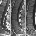
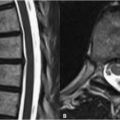
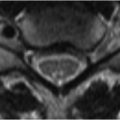
44 Disk Herniations Pathologic extensions of the intervertebral disk beyond the vertebral end plate margin are categorized as bulges, protrusions, extrusions, or free fragments. A disk bulge results from laxity of and tears within the annulus fibrosis that allow nonfocal extension of the nucleus pulposus posteriorly, with the posterior disk margin forming a smooth, curvilinear contour. Disk bulges are broad-based, circumferential, and may narrow the spinal canal and neural foramen. A disk protrusion, in distinction, is a focal extension or herniation of disk material beyond the posterior end plate margin. Disk protrusions and extrusions are similar but distinct entities, extrusion referring to a protrusion of the nucleus pulposus in which no intact annular fibers remain. This distinction cannot reliably be made on MRI, and thus the term disk protrusion is generally reserved for a small herniation and extrusion for a large herniation. It is imperative to distinguish whether a disk herniation is central, paracentral, or foraminal in location. Figures 44.1A and 44.1B demonstrate a disk extrusion at the L4–L5 level on sagittal T2WI and axial T1WI, respectively. Herniations at L4–L5 and L5–S1 comprise 90% of lumbar herniations. This particular extrusion migrates inferiorly on (A
![]()
Stay updated, free articles. Join our Telegram channel

Full access? Get Clinical Tree


