Tendons
Tibialis Anterior
Anatomy
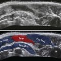
The tibialis anterior tendon is the strongest of the anterior ankle tendons. Its parent muscle arises from the upper two-thirds of the lateral aspect of the tibia. It has a high musculotendinous junction and a strong distal tendon, which is constrained by a transverse superior and an oblique inferior retinacular band. The relationship of tibialis anterior to the retinacula is complex. The superior retinaculum is band-like and lies at the level of the distal tibia below the musculotendinous junction ( Fig. 25.1 ). This retinaculum represents the anterior portion of a circumferential band of tissue that includes the tarsal retinaculum, overriding the tarsal tunnel, and the superior peroneal retinaculum laterally. In cross section, there may be two components, superficial and deep, with the tibialis anterior tendon passing in a tunnel between them. This tunnel may also separate tibialis anterior from the other extensor tendons. The oblique inferior retinaculum comprises two bands, a superomedial and an inframedial, which unite to form a single lateral attachment. The tibialis anterior tendon passes deep to the oblique inferomedial ligament just prior to its insertion into the medial cuneiform. The tendon insertion is quite far medial onto a facet of the medial cuneiform and base of the first metatarsal.
There are two areas where tibialis anterior is susceptible to injury. The first is between the retinacula, where tendon rupture occurs, and the second is at the insertion, where tendinopathy and enthesopathy are more common.

Tenosynovitis and Tendinopathy
Tendinopathy of the tibialis anterior tendon is rare, but may occur as an overuse injury. Repetitive foot dorsiflexion during sports such as running, skiing, cycling, and mountain climbing has been implicated in the aetiology of the disorder. On other occasions, footwear has been blamed and, in particular, direct irritation from the upper edge of the shoes or boots may be the cause.
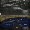
The earliest changes are found around the tendon where it abuts the retinacula. Fluid may gather around the tendon ( Fig. 25.2 ), though it will be constrained in the area where the retinaculum compresses it ( Fig. 25.3 ). With progression, the tendon itself becomes involved, leading to thickening and decreased reflectivity ( Fig. 25.4 ).
| In some cases, the tibialis anterior tendon synovial sheath may be uninvolved, but there is thickening of the retinaculum, leading to a type of stenosing tenosynovitis ( Fig. 25.5 ). |


Tibialis Anterior Enthesopathy
| Isolated enthesopathy may involve the distal portion of tibialis anterior tendon at its insertion. |

Tendon Rupture
The commonest site for tibialis anterior rupture is in the space between the superior and the inferior retinaculum. Attention has been drawn to an area of poor vascularity within the tendon in this area. Rupture may also occur beneath the oblique superomedial limb of the inferior extensor retinaculum. A further site of tendon injury is at its insertion into the medial cuneiform. It is not uncommon to identify enthesophyte formation in this location that, if marked, may abrade the tendon, leading to rupture.
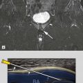
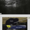
Symptoms of anterior tibialis tendon rupture are often subtle. This is because the loss of dorsiflexion resulting from the injury may be compensated by other tendons. A common presentation is with an anterior mass ( Fig. 25.7 ), often sufficiently prominent to lead to concerns that a soft tissue tumour is present ( Fig. 25.8 ). The mass is due to a combination of fibrosis in the extensor retinaculum and the torn lax tendon. Other ultrasound findings include a tapering appearance to the tendon end ( Fig. 25.9 ) and retraction ( Fig. 25.10 ).