Disorders of the Elbow
Posterior
Olecranon Bursitis
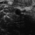
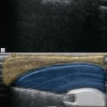
One of the commonest causes of localized pain in the posterior elbow is olecranon bursitis. This is usually a clinical diagnosis as bursa enlargement is easily palpable in the typical location above the olecranon. Occasionally in patients with large limbs, subtle enlargement may be difficult to detect clinically and imaging may be helpful in these cases (Fig. 9.1).
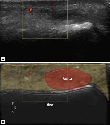
Figure 9.1 Sagittal section of posterior elbow. An ill-defined predominantly low-reflective mass underlies the proximal olecranon. Enlargement of the olecranon bursa is one of the commonest masses around the elbow.
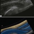
The cause is usually due to repetitive friction as the characteristic location of the olecranon bursa makes it particularly susceptible to trauma, leading to haemorrhage within the bursa. Conditions associated with inflammatory synovitis also commonly affect the bursa (Fig. 9.2). Septic bursitis, particularly related to penetrating injuries, also occurs.
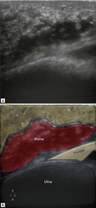
Figure 9.2 Sagittal posterior elbow and poorly defined fluid collection underlying the olecranon. Another example of olecranon bursitis.
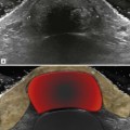
The wall may be thick or thin depending on the degree of associated synovitis and, if synovial thickening is present, it may be active on Doppler or inactive and representing fibrous pannus.