Abstract
Duplication of the renal collecting system is recognized in approximately 1 : 4000 pregnancies and is more frequent in females. It disproportionately affects the left kidney, and is bilateral in 15% to 20% of cases. Ultrasound (US) may show dilatation of one or both renal pelves, with an intervening band of renal tissue. Ureteral dilatation may be evident, and there may be a ureterocele visible in the bladder. The Weigert-Meyer rule describes the relationship of the ureters. The upper pole ureter inserts medially and inferiorly to the lower pole ureter, with obstruction of the upper pole moiety secondary to a ureterocele. The lower pole ureter inserts laterally in the bladder trigone and has a shortened intravesical segment that leads to reflux. When a diagnosis of a duplicated collecting system is made prenatally, neonatal evaluation is important, as these individuals are prone to reflux, obstruction, and urinary tract infection.
Keywords
duplex kidney, ureterocele, Weigert-Meyer rule
Introduction
Duplication of the renal collecting system, also termed duplex kidney, is one of the few renal anomalies more common in females. Duplex kidneys have an upper pole and a lower pole, called moieties, each drained by a ureter. Approximately one-third of the duplex kidney is drained by the upper pole ureter, with two-thirds drained by the lower pole ureter.
Many individuals with duplex kidneys are asymptomatic and have no impairment of renal function. Those who come to attention prenatally or postnatally usually have a complication related to abnormal implantation of one or both ureters. The upper pole ureter may form a ureterocele within the bladder that causes obstruction, resulting in little or no function of the upper pole moiety. The lower pole ureter may have a short intravesical segment that results in vesicoureteral reflux (VUR). Duplex systems that have these complications also have ultrasound (US) findings that facilitate their prenatal detection. Clinically significant duplication of the collecting system may manifest prenatally with upper pole hydronephrosis and dilatation of the upper pole ureter, lower pole hydronephrosis, or a ureterocele within the bladder.
Disorder
Definition
Duplication of the renal collecting system is a condition in which a kidney is divided into separate upper and lower pole moieties, each drained by a ureter. If the duplication is complete , the upper and lower pole ureters drain separately. If the duplication is partial, the ureters fuse to form a bifid ureter before emptying into the bladder. Complete duplication is often associated with a ureterocele, which is a cystic dilatation of the distal end of the ureter, usually within the bladder but occasionally at other sites along the genitourinary tract.
Prevalence and Epidemiology
The prevalence of duplicated collecting systems is difficult to estimate accurately because most cases are not identified either prenatally or in childhood. Prenatally detected renal duplication occurs in approximately 1 : 3000 to 1 : 5000 births, based on data from the 1990s. In autopsy studies, the prevalence of a duplicated system (including partial duplication) is approximately 1 : 125.
Renal duplication is twice as common in females, and 80% of cases complicated by a ureterocele occur in females. The duplication is bilateral in 15% to 20%. Unilateral cases are more likely to affect the left kidney. Duplication is generally considered to be a sporadic anomaly, although an increased incidence has been described in some families, suggesting autosomal dominant inheritance with incomplete penetrance and variable expressivity.
Etiology, Pathophysiology, and Embryology
Complete duplication occurs when an extra ureteric bud arises from the mesonephric (Wolffian) duct and fuses with the metanephric mesenchyme at a separate site from the original bud. The ureteric bud closest to the urogenital sinus gives rise to the lower pole ureter, and the cephalad bud gives rise to the upper pole ureter. Partial duplication occurs when a single ureteric bud divides before fusing with the metanephric mesenchyme.
The anatomic relationship of the upper and lower pole ureters as they enter the bladder is called the Weigert-Meyer rule . The upper pole ureter crosses the lower pole ureter and inserts medially and distally (inferiorly) to the lower pole ureter, either ectopically in the bladder or occasionally elsewhere along the genitourinary tract. Sites include the uterus, vagina, urethra, ejaculatory ducts, vas deferens, epididymis, or seminal vesicles. The upper pole ureter often develops a ureterocele, a cystic dilatation of the intravesical portion of the ureter, with narrowing of the ureteral orifice. In the setting of obstruction from a ureterocele in a duplex system, two-thirds of individuals have poor function of the upper pole moiety. The lower pole ureter drains laterally in the bladder trigone. The lower pole ureter has an intravesical portion that is shorter than normal, which may result in VUR and eventual cystic renal dysplasia.
Manifestations of Disease
Clinical Presentation
Prenatal diagnosis of a duplicated collecting system is based on US findings, as discussed subsequently. Most cases do not manifest prenatally, and many remain undiagnosed throughout life, with no renal impairment. Affected children may present with urinary tract infection secondary to reflux. Less commonly, individuals may be found to have a duplex system during evaluation for hypertension or renal insufficiency. Rarely, symptoms may result from implantation of the upper pole ureter in a location other than the bladder, such as incontinence from ureteral implantation in the vagina.
Imaging Technique and Findings
Ultrasound.
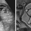
The characteristic US finding is dilatation of the upper and lower pole pelves, with an intervening band of renal tissue ( Fig. 13.1 ). The degree of hydronephrosis affecting each moiety can range from simple dilatation of the pelvis to marked calyceal dilatation with thinning of the renal cortex (see Fig. 13.1 ). Visualization of the intervening band of tissue differentiates renal duplication from other causes of renal pelvis dilatation (see Chapter 12 ). With improvements in ultrasound technology, it may be possible to identify a duplex kidney when neither moiety is dilated – which is termed an uncomplicated duplication ( Fig. 13.2 ). Identification of two renal arteries supplying the kidney further supports the diagnosis (see Fig. 13.2 ). The length of the kidney often exceeds the 95th percentile (see Fig. 13.2 and Table 9.1 ). Whenever renal pelvis dilatation is encountered, it is important to image both kidneys in the sagittal and transverse planes, so that a duplex system is not missed. Eccentric location of a dilated single renal pelvis (high or low in the kidney) warrants a careful evaluation of the renal parenchyma, the ureter exiting the pelvis, and the bladder ( Fig. 13.3 ).

Related posts:


Stay updated, free articles. Join our Telegram channel

Full access? Get Clinical Tree


