
In sport, pain is part of the deal. Every training session leaves its small souvenirs: heavy legs, stiff back, a bruise here and there. But there is a big difference between “I’m tired” and “something is broken”. The problem is that, in the heat of competition, many players pretend not to feel that difference. They tape the knee, swallow a pill, say a quick prayer and go back on the pitch. That’s how a small injury quietly grows into a big problem.
Modern imaging is the flashlight in this dark room. X-ray, CT and MRI are the tools that show what the naked eye can’t see, long before the body sends the final invoice in the form of chronic pain or a ruined season.
Time: the most expensive medicine
One truth every coach understands: what you ignore today, you pay double for tomorrow. In sports medicine it works the same way. A muscle tear scanned in the first 48 hours can be graded, treated and monitored. The same tear, ignored for weeks, can heal poorly and become a weak spot that rips again every time the player accelerates.
Early imaging does three essential things:
- Confirms what the injury actually is (not just what it feels like).
- Shows how big the damage is and which structures are involved.
- Helps doctors design a realistic return-to-play plan instead of guessing.
For professionals, a clear scan can be the difference between missing three games or missing a whole year. For amateurs, it decides whether they will still be able to play with their children in ten years or walk with a limp before forty.
X-ray: the first gatekeeper
X-ray is the old warrior of medical imaging. It doesn’t show every detail, but it’s fast, cheap and perfect for detecting fractures, dislocations and certain bone diseases. When a player falls awkwardly, gets kicked in the shin or lands badly after a jump, X-ray is usually the first stop.
It shows whether that painful ankle is “just” a sprain or actually a small fracture around the joint. It reveals stress fractures in a runner’s foot, who thought it was only fatigue. And in contact sports, it helps check for damage around the shoulder, ribs and spine after heavy collisions.
The key point: without a simple X-ray, people often walk on broken bones, waiting for them to “settle”. They do settle – in the wrong position. Later, the same player complains that the joint “never felt the same again”, not realizing the bad healing started the day they refused the first scan.
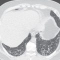
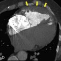
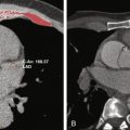
CT scan: zooming in on complex injuries
CT (computed tomography) is like X-ray with a magnifying glass and a brain. It creates cross-section images of the body, making complex areas much clearer: the spine, the face, the small bones of the wrist and ankle, or joints damaged in multiple places.
In sports, CT is beneficial after severe trauma, such as a high-speed collision, a fall from height, or a player presenting to the emergency room after a road accident. Doctors use CT to map fractures in 3D, check for internal bleeding and spot small bone fragments that could later irritate tendons or cartilage.
For example, a goalkeeper who hits the post with his head might need CT to rule out a skull fracture or bleeding in the brain. Without that scan, he might leave the hospital with “just a headache”, go back to training too soon and risk something much worse.
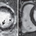
MRI: seeing the invisible
If X-ray and CT are good with bones, MRI is the master of soft tissues: ligaments, tendons, cartilage, muscles, nerves and the brain itself. It is the tool that tells the truth about the cruciate ligament, the meniscus, the Achilles tendon or the hamstring that keeps “pulling” again and again.
MRI shows precisely where the tissue is torn, how deep the tear is and whether there is hidden damage surrounding it. In knee injuries, it can reveal that a simple twist has actually injured three different structures at once. In head injuries, it helps assess concussion, swelling and subtle changes in the brain.
Because of this detail, MRI is the gold standard for deciding whether a player needs surgery or can recover with physiotherapy. It also helps measure healing over time, so the doctor can say: “Your scan is clean, we can start high-intensity work,” instead of relying on wishful thinking and the player’s optimism.
From street pitch to national stadium: different risks, same body
Whether someone plays barefoot on a dusty community field or in a perfect stadium with a GPS vest, the body works with the same muscles, the same bones and the same fragile ligaments. The difference is access. Top clubs have their players scanned the same day they get injured. Ordinary people, and even semi-pro athletes, hesitate. They worry about cost, travel, missing work, or they simply hope that time alone will fix everything.
The result is predictable: meniscus tears turning into early arthritis, untreated fractures deforming the joint, repeated concussions stacking up until simple noises cause headaches. On the surface the person “just got old,” but the real story is delayed diagnosis. Early imaging is not a luxury for rich stars; it is basic maintenance for anyone who wants their body to last.
Odds, injuries and the lure of the betting slip
On match day, the drama of injuries doesn’t only live in the physio room. It lives in the odds too. The moment a star striker limps off or a key defender clutches his hamstring, you can see the live numbers change on screens. Sports betting companies adjust quickly, because they know that one MRI result can reshape a whole season.
Many fans follow that dance between medicine and odds through platforms where they place small slips before the game or react in-play. For some, checking a service ethio bet is as normal as checking the weather: they look at the injury news, read how long a player will be out, and decide whether to back the favourite or search for value in the underdog. Responsible brands try to educate users about team news, injuries and rotation, so that betting becomes a matter of informed decisions rather than blind guesses. When people understand how crucial early scans are for a player’s recovery, they also understand why today’s “easy” fixture might suddenly be much more complicated.
Smart athletes, smart bettors: respecting the body
Injuries don’t only shape the line-up; they shape how people think about risk. A bettor who has lost money because a half-fit player broke down after 20 minutes learns a hard lesson: ignoring medical realities is expensive. That is why many fans now read medical updates with the same attention they give to tactics. If the club’s doctor says the winger returned “ahead of schedule”, the careful bettor hears: “Maybe the risk is still high.”
Platforms associated with serious operators, including melbet ethiopia, try to position themselves on the side of information, not illusion. They publish previews that talk honestly about injuries, suspensions and recovery times. They also push messages about responsible gambling: setting limits, treating betting as entertainment, not as a salary. Just as a professional must know when to rest a sore knee, a fan must know when to step away from the odds for a while. Respecting the body and respecting your bankroll are two versions of the same wisdom.
What early imaging really protects
It’s easy to think of scans as cold machines in a hospital basement. But behind every X-ray, CT or MRI there is a human story. Early imaging protects:
- Careers, by catching damage before it becomes permanent.
- Families, by keeping a parent able to work and play, not just sit on the sidelines.
- Teams, by avoiding the gamble of throwing half-fit players into high-risk games.
- Even bettors, by making the picture more transparent and reducing the chaos around injuries.
A wise person doesn’t wait for the roof to fall before checking the leaks. In sport, the body is that roof. Pain is the drip from the ceiling. Imaging is the ladder and the flashlight. Use it early, and you may only need a small repair. Wait too long, and one day the whole house will come down – no matter how strong you thought it was.
Related posts:


Stay updated, free articles. Join our Telegram channel

Full access? Get Clinical Tree


