Fig. 13.1
Typical skin rashes of a macaque infected by EBOV-Z. (a) Diffusive bleeding spots on the right arm of the macaque. (b) Multiple bleeding spots scattering in the inguinal region of the macaque (Reproduced with permission from Thomas W, et al. AJP 2003;163(6):2347)
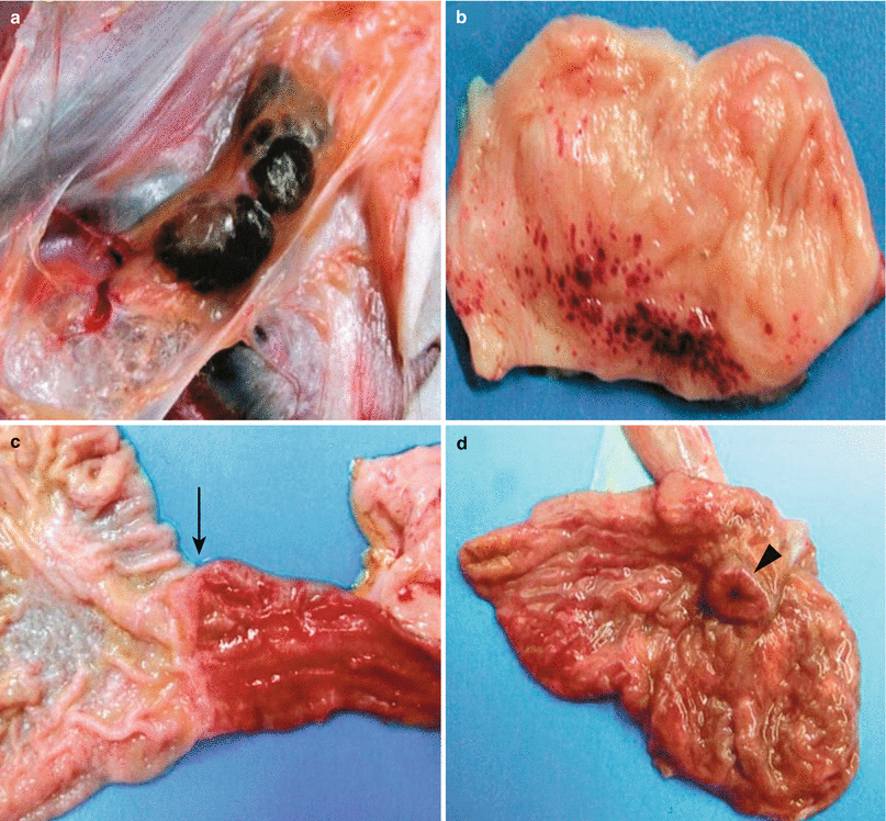
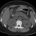
Fig. 13.2
Autopsy specimen of the macaque infected by EBOV-Z. (a) Enlarged lymph nodes from the inguinal region of the macaque, with bleeding and congestion. (b) Multiple focal bleeding spots in the adrenal gland. (c) Duodenal congestion (pointed by arrow). (d) Congestion at the ileocecal junction (pointed by arrowhead) (Reproduced with permission from Thomas W, et al. AJP 2003;163(6):2347)
13.4 Clinical Symptoms and Signs
The incubation period of EHF generally lasts for 2–21 days, commonly 5–12 days. The cases of EBOV infection can be asymptomatic or have mild symptoms. Non-critical cases gradually recover 2 weeks after the onset. The typical cases with EHF have an acute onset, with clinical manifestations of high fever, aversion to cold, headaches, muscle and joint pain (commonly major joints and lumbosacral region), vomiting, conjunctival congestion, and relatively slow pulse. Gastrointestinal symptoms occur 2–3 days after the onset, characterized by diarrhea (firstly watery and following dark) along with vomiting and angina. The cases develop into the climax stage 4–5 days after the onset, characterized by conscious change such as delirium and drowsiness. In serious cases, hemoptysis and bleeding in the nose, mouth, subconjunctiva, gastrointestinal tract, vagina, and skin as well as hematuria can be found several days after the onset. The bleeding peaks at day 10 after the onset. Due to necrosis and decomposition of the organs, patients continually vomit the necrotic tissue via their mouth. Above 50 % of the patients have serious bleeding, and death occurs due to bleeding as well as hepatic and renal failure. The prominent manifestations include hypotension, shock, and facial edema, DIC, as well as electrolytes and acid-base balance disturbances. After infection of EBOV by pregnant women, uterine bleeding and miscarrying occur. In the death cases, 90 % have its occurrence within 12 days after the onset. The early symptoms after the onset of EHF are deceptive and tend to be misdiagnosed as common cold, flu, measles, cholera, typhoid fever, malaria, and HIV/AIDS that are common in Africa. Thus, its early diagnosis is difficult to be defined. However, its definitive diagnosis after various bleedings is too late to save the life.
13.5 EHF Related Complications
Complications in the acute stage include myocarditis and bacterial pneumonia. In not serious cases, patients gradually recover in 2 weeks after the onset. Most patients have asymmetrical joint pain that may be migratory that mainly involves major joints. Some patients show delayed symptoms, including myalgia, fatigue, suppurative parotitis, hearing loss or tinnitus, conjunctivitis, unilateral blindness, and uveitis. Due to the long-term survival of the virus in the seminal fluid, delayed diseases such as testitis and testicular atrophy can be induced. At day 5–7 during the course of the disease, measles-like macula may occur that is common on the shoulders, palms, and soles. The macula is absent with desquamation after several days. Some patients may have long-term residual skin changes.
13.6 Diagnostic Examinations
Laboratory tests for the diagnosis of EHF mainly include etiological examination, serologic test, electron microscopy, and virus isolation. The serologic test and virus isolation are more commonly applied in clinical practice.
13.6.1 Laboratory Tests
13.6.1.1 Routine Tests
Routine Blood Test
In the early stage, WBC count decreases that increases since day 7 after the onset of disease, along with heteromorphic lymphocytes and possibly thrombocytopenia.
Routine Urine Test
In the early stage, proteinuria can be found.
Biochemical Test
By biochemical test, AST and ALT levels increase and the increase amplitude of AST is greater than ALT.
13.6.1.2 Serologic Test
After EBOV infection by human, specific antibody can be detected in the serum. IgM antibody soon reaches its peak 1 week after the onset, while IgG antibody begins to increase 1 month after the onset. In the early stage of the disease, indirect immunofluorescence and enzyme-linked immunosorbent assay can be applied to detect the serum IgM for definitive diagnosis. Otherwise, in the convalescence stage of the disease, the finding of four times increase of antibody in double sera sample can define the diagnosis. Other tests such as dot blot have also been reported as the diagnostic examination of EHF.
13.6.1.3 Etiological Examination
EBOV is highly dangerous and relevant experiments should be conducted in BSL-4 laboratories.
Virus Antigen Detection
During the course of EBOV, high-titer viremia occurs. Therefore, the virus antigen can be detected in serum by ELISA. Immunofluorescence is also widely applied to detect the virus antigen in the liver and spleen of infected animals.
Virus Isolation
The specimens of blood, serum, plasma, and seminal fluid can be collected during the acute stage of the disease to be inoculated into Vero cells. After culture, the ultrathin sections can be observed under an electron microscope to detect the typical filament-like virus particle in the cytoplasm. The virus can also be isolated by intracerebral inoculation of neonatal rats with following electron microscopy or serological test. The diagnosis can be defined based on successful detection of the virus.
Related posts:

Stay updated, free articles. Join our Telegram channel

Full access? Get Clinical Tree


