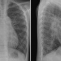
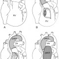
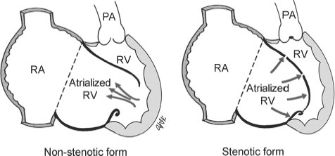
14 Ebstein’s Malformation and Other Forms of Congenital Tricuspid Regurgitation Fig. 14.1 Ebstein’s malformation of the right tricuspid valve. PA, pulmonary artery; RA, right atrium; RV, right ventricle.
Definition and Classification
 Any abnormality of the tricuspid valve resulting in congenital tricuspid regurgitation of varying severity
 Ebstein’s malformation (Fig. 14.1) results from downward displacement of tricuspid valve tissue attachment below the normal annulus and dividing the right ventricle into a proximal atrialized portion and a distal functional right ventricle. Displaced attachment typically involves the septal and posterior leaflets of the valve. The anterior leaflet does not usually show displaced attachment but is usually large and sail-like. As the leaflet edges are not aligned properly, varying degree of tricuspid regurgitation (Fig. 14.1, arrows) characterizes this malformation. Occasionally, the enlarged anterior leaflet may fuse with the displaced leaflets to form a sac of atrialized right ventricle, causing tricuspid stenosis or atresia. The latter form can be associated with functional or anatomic pulmonary atresia.
 Similar hemodynamic consequences in other congenital tricuspid valve malformations, with variable dysplasia having deficient leaflets, extra leaflets, unguarded tricuspid orifice, and isolated clefts
 Transient tricuspid insufficiency in infants caused by myocardial dysfunction most often secondary to pulmonary hypertension
 Isolated giant right atrial aneurysm a rare condition with idiopathic dilatation of the right atrium associated with tricuspid annular dilation and tricuspid regurgitation
 Associated lesions include congenitally corrected transposition, a variety of left heart lesions, and conduction anomalies
Pathophysiology
Stay updated, free articles. Join our Telegram channel

Full access? Get Clinical Tree


