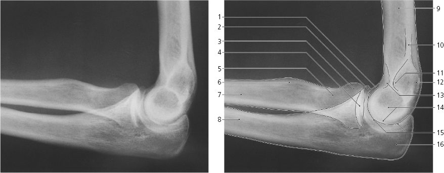
Olecranon fossa, and coronoid fossa (superimposed)
Lateral epicondyle
Capitulum
Humeroradial joint
Head of radius
Neck of radius
Shaft of radius
Medial supracondylar ridge
Medial epicondyle
Olecranon
Trochlea
Coronoid process
Articular circumference of radius
Radial tuberosity
Shaft of ulna
Elbow, lateral X-ray
Capitulum
Coronoid process
Head of radius
Articular fovea of radius
Neck of radius
Radial tuberosity
Shaft of radius
Shaft of ulna
Shaft of humerus
Medial supracondylar ridge
Olecranon fossa
Medial epicondyle
Coronoid fossa
Trochlea
Humero-ulnar joint
Olecranon
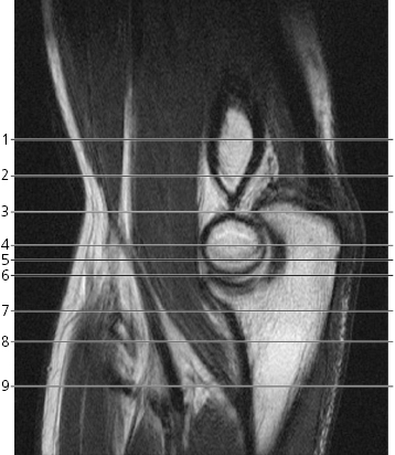
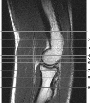
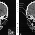
Scout views of elbow
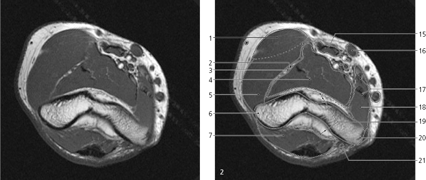
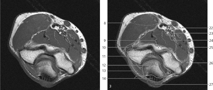
Lines #1–9 indicate planes of sectioning in the following axial MR series. Interpretation of the scout images can be found in the sagittal series, page 73–74, image #1 and #3. Note that the radial artery in this series has branched off from the brachial artery before reaching the cubital fossa. The frequency of this variation is about 15%. The artery termed “brachial artery” below might as well be termed “ulnar artery”. However, it takes up the position of the brachial artery. The forearm is pronated.Elbow, axial MR
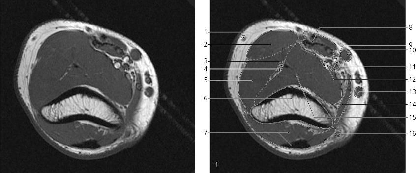
Cephalic vein →
Brachioradialis →
Radial nerve, superficial branch →
Radial nerve, deep branch →
Extensor carpi radialis longus →
Humerus →
Triceps brachii, muscle and tendon →
Biceps brachii →
Radial artery (high division) with comitant veins →
Median cubital vein →
Median nerve →
Brachial artery with comitant veins →
Basilic vein →
Pronator teres (humeral head) →
Brachialis muscle →
Ulnar nerve →
Elbow, axial MR
Only gold members can continue reading. Log In or Register to continue