Imaging variants of the elbow and pitfalls can be disconcerting and can lead to diagnostic mistakes. Inhomogeneities in the magnetic field and coil position can result in signal changes that may simulate abnormality. Bone signal and morphology variants, such as the islands of red marrow and the pseudodefect of the capitellum and intraarticular inclusions such as plicae, may be mistaken for abnormal findings. Variations of the distal biceps and triceps tendons and different aspects of the ligaments and their insertions, as well as nonpathologic signal and width changes in the ulnar nerve, are other examples of common pitfalls in magnetic resonance imaging of the elbow.
Recognition of imaging variants has been appropriately stressed since not long after the first radiograph was performed. These variants are most disconcerting on more advanced imaging and in those parts of the body that are less imaged. Both of these qualifiers apply to magnetic resonance (MR) imaging of the elbow.
MR imaging field inhomogeneity
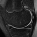
Inhomogeneities in the magnetic field and coil positioning can create areas of variable fat saturation or increased signal in tissues because of varying proximity to the coil, which may simulate bone marrow edema in the olecranon, bursitis in the adjacent subcutaneous tissues, or even tendinopathy in the tendinous insertions in the epicondyles. As the elbow is often imaged away from the isocenter, inhomogeneous fat suppression is a particular problem. In the authors’ experience, fat suppression creates the most diagnostic difficulties in the distal triceps and in the region of the olecranon bursa ( Fig. 1 ).