Endocrine, Aerodigestive Tract, and Breast Tumors


Endocrine, Aerodigestive Tract, and Breast Tumors


Line Claude

Cécile Nozières


ADRENOCORTICAL CARCINOMA


Histology

Adrenocortical carcinoma (ACC) is an aggressive cancer originating in the cortex of the adrenal gland. This chapter focuses on corticosurrenaloma. Different types of tumors exist (myxoid adrenocortical carcinoma, carcinosarcoma, oncocytic adrenocortical carcinoma, and clear cell adrenal carcinoma), but are very rarely described in children.


Epidemiology

ACC is rare around the world (0.2% of child cancer) (1, 2, 3, 4, 5, 6, 7), except in Southern Brazil (3.5 cases/million children younger than 15 years) (8) due to p53 mutation. The frequency is dependent on the age: 0.4 per million before 4 years and decreases to 0.1 per million after 10 years (9). Sex ratio is 1:5.3 before 4 years and 1:0.8 after 4 years (8).


Genetics

Principal genetic abnormalities associated with ACC are summarized in Table 16.1 (10).


Clinical Aspects

Virilization (pubic hair, clitoromegaly or phallomegaly, acne, deep voice, and hirsutism) is the most common sign of ACC. In the large Brazilian experience, more than 90% of the patients had pubic hair, and more than 80% of girls had clitoris hypertrophy. Other frequent signs are abdominal pain in 50% and Cushing syndrome in 30% (hypertension, centripetal fat distribution, moon face, buffalo hump of the neck, accelerated growth velocity, weight gain, …) (Fig. 16.1).


Table 16.1 Constitutional Genetic Abnormalities Associated with Adrenal Cortical Tumours (ACT)


Condition


Tumor Types


Li-Fraumeni syndromes and other germ-line p53 mutations


Adenomas/carcinomas


Hemihypertrophy


Adenomas/carcinomas


Beckwith-Wiedemann syndrome


Adenomas/carcinomas


Carney complex


Primary pigmented nodular adrenocortical disease


Congenital adrenal hyperplasia


Adenomas carcinomas (very rare)


Multiple endocrine neoplasia I


Adenomas carcinomas


From Ribeiro RC, Figueiredo B. Childhood adrenocortical tumours. Eur J Cancer. 2004;40(8):1117-1126, with permission.


Biology

Because the tumor cells may secrete a number of adrenal hormones either alone or in combination (androgens, glucocorticoids, mineralocorticoids, and estrogens), it is possible to establish an endocrine profile of most pediatric ACC. Urinary 17-ketosteroids (17-KS) and 17-OH steroids are elevated in more than 90% of the cases (8).


Molecular Biology

In adult series, Ki-67 marker proliferation, p53 mutation, mdm-2 mutation, and p21 mutation have been identified as associated with ACC but no specific pattern is known. While tumor cell proliferation (Ki-67) correlates with mitotic activity and morphologic index, tumor morphology remains better predictor of metastatic risk (11).


Prognostic Factor

The distinction between benign and malignant ACC is made on cytologic abnormality, tumor weight, and presence of metastases (12,13). Some lesions are frankly malignant on microscopic examination. For those of borderline histologic appearance, one should consider resectability, the extent of capsular invasion, adherence to surrounding structures, the presence of aberrant vessels on angiography, tumor size, and the presence or absence of metastases to distinguish benign from malignant lesions. Well-encapsulated and easily excised tumors may be cured by surgery alone. In the remaining resected but more locally advanced cases, there is a substantial risk of local relapse and distant metastases (7,14,15).


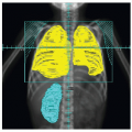
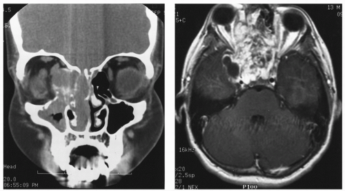
Figure 16.1 Coronal computed tomography (left) and axial magnetic resonance imaging (MRI) (right) of a 13-year-old boy with headaches, diplopia, and right proptosis. The mass-produced destruction of the ethmoid air cells and superior turbinates, mass effect in the right orbit, compression of the right cavernous sinus, displacement of the right internal carotid artery, and displacement of the pituitary. A biopsy proved the diagnosis of juvenile nasopharyngeal angiofibroma. Three-dimensional treatment planning was used to prepare a four-field noncoplanar external beam radiotherapy treatment technique using rigid head immobilization. A dose of 36 Gy at 2 Gy per fraction was administered. One and one-half years after irradiation the symptoms had significantly improved and the mass was smaller on physical examination and MRI. Shortly thereafter the symptoms recurred, and the MRI demonstrated regrowth of the angiofibroma, within and outside the irradiated field. Interferon was administered as antiangiogenesis agent.

While stages are well defined in adults (McFarlane classification (4)), classification is less clear in childhood. Michalkiewicz et al. proposed a classification based on more than 250 patients (9). Factors independently associated with good prognosis in terms of survival include Stage I tumor (tumor completely excised, tumor weight <200 g, and absence of metastasis), virilization alone, age <4 years (9).


Imagery

Imaging studies are of great importance for surgical planning and disease staging. Magnetic resonance imaging (MRI) and tomodensitometry (TDM) are necessary to determine the size of tumor, to detect invasion of local structures, metastasis, and vessels involvement (vena cava, for example).

Due to frequent liver and lung metastasis, TDM is recommended. Brain and bone metastases are rare but possible; technetium bone scan and/or brain MRI should be performed in case of observed symptoms. Positron emission tomography (PET) scan has not been extensively studied, but in a very small series in adults, it can yield additional information in defining tumor metabolic activity and necrosis, and in the earlier detection of metastases in comparison with computed tomography (CT) (16).


Treatment


Surgery

For localized disease, surgery is usually recommended although it is the most important procedure in successful treatment of ACC (8). Tumor friability needs a bloc resection to avoid tumor spillage and rupture of the capsule and to compromise prognosis. Therefore, transabdominal approach is necessary.


Chemotherapy

Patients with residual disease after surgery or with metastatic disease usually receive chemotherapy using different regimen, including mitotane, cisplatin, etoposide, doxorubicin, with poor results (17). At one time, mitotane was considered to be the drug of choice. This drug, an isomer of the insecticide DDD, has adrenolytic activity. It has been used in neoadjuvant to treat advanced metastatic ACC in cases of inoperable tumors, in adjuvant situation for patients at high risk to relapse, and to control symptoms associated with production of adrenal hormones (10). Administration of this drug is associated with toxic effects such as nausea, vomiting, renal and hepatic dysfunction, and neurologic alterations. As in adults series, response rates to mitotane are heterogeneous (0%-56%), highly dependent of serum therapeutic level (14 to 20 µg/mL (17). Association with others drugs, doxorubicin, cisplatin, and 5-fluorouracil (FU), was possible, without evident benefice in tumor response.


Radiotherapy

Radiotherapy (RT) has often been considered ineffective for treatment of ACC. However, old clinical reports have described tumor response rates up to 42% (18), which indicate that ACC is not resistant to RT.

The only series reported with children is Magee’s study (12). Fifteen patients including five children were treated for ACC. Nine of them had postoperative irradiation. Three were girls who presented under the age of 2 years with hormonally active tumors and who survived for more than 10 years after gross total tumor resection followed by 30 Gy in 4 weeks. Two of these three patients died of second malignancies arising in the irradiated field. The high frequency of second
primary malignancies is observed in other studies, and it points out the need for close surveillance especially after RT (19).

Recommendation of an international consensus conference was published in 2005 for adults without any data for children. Radiation therapy is recommended in the treatment of bone, brain, and other metastases. It is also recommended in the treatment of symptomatic local recurrences. Concerning adjuvant situations, it should be discussed in case of incomplete local resection. There is no recommendation concerning adjuvant RT in cases of complete resection, and no major evidence to support it (20). However, a more recent study demonstrated the efficiency of adjuvant RT of the tumor bed (± bilateral paraaortic lymph node in seven patients due to lymph node involvement) in a retrospective cohort. Fourteen patients (eight in complete resections, two marginal resections, and four unknown) received adjuvant RT (median dose 50.4 Gy, range [41.4-56]. 1.8-2 Gy per fraction, beginning within 8 weeks after surgery in most patients) while 14 were in the control group. Local control was significantly better in the radiation group: 79% at 5 years versus 12% at 5 years without RT (p < 0.01), but without impact on survival (21) (Fig. 16.2).


Follow-Up

Consensus recommends hormonal monitoring every 2 months in the first years, followed by monitoring every 4 months in the second years and every 6 months from then on (20). Imaging studies are indicated only in the presence of hormonal abnormalities in case of functional ACC.


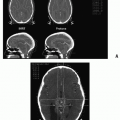
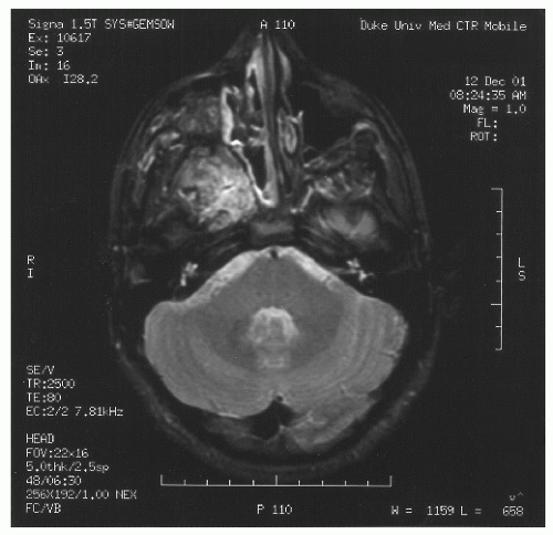
Figure 16.2 A 14-year-old boy presented to the radiation oncology department after multiple surgical and embolization procedures intended to remove and devascularize a juvenile nasopharyngeal angiofibroma. Detailed three-dimensional mapping of the tumor was performed using magnetic resonance imaging accompanied by careful fiber-optic examination. The mass was treated, with a margin, with a mixture of 6- and 15-MV photons to a cumulative dose of 40 Gy. Over the subsequent 2 years, striking regression of the mass was seen. Before RT, the child had been subjected to repeated episodes of bleeding, which ceased after RT.


Results

In children, considering all the stages, 5 years of overall survival (OS) ranges from 34% to 54%. Event-free survival (EFS) and OS are very close (9). Stages (quality of resection, tumor weight, and metastasis), age, and virilization are factors significantly associated with survival. In patients with stages III or IV, OS is from 0% to 20% at 5 years. Five years of OS for stages I and II is 80%-100% and 30%-50%, respectively (9).


PHEOCHROMOCYTOMA: PARAGANGLIOMA


Definition

These tumors arise from the chromaffin cells in the sympatheticadrenal system (22, 23, 24). Pheochromocytomas (PHs) are developed from medullary gland, whereas paragangliomas (PGLs) come from extramedullary chromaffin cells (Zuckerkandl organ, bladder, kidney hilar, posterior mediastinum, pericarditis, head, and neck) (25). Adrenal medullary PHs produce epinephrine and norepinephrine, whereas most PGLs (about 10% of cases) produce only norepinephrine. Some of these tumors are nonsecretor. The risk of malignancy is higher for PGL than for PH (26).


Histology

As with many other neuroendocrine tumors, distinction between benign and malignant tumors is difficult. The histologic scaling system “Pheochromocytoma of the Adrenal Gland Scaled Score” uses a range of histologic criteria (vascular and capsular invasion, necrosis, and increased mitotic figures) to determine aggressive behavior, but it cannot predict malignancy. Histologic markers (Ki67, p53) may be indicative of a malignant disease, but none has been shown to be a prognostic marker (27). Malignancy is defined retrospectively by presence of metastases (25).


Epidemiology

The incidence of PH and PGL in the pediatric population is rare, accounting for <1% of childhood cancer, with an incidence of 2 per million (28, 29, 30). Two third of pediatric cases occur in boys. The incidence of malignant PH range depends on genetic background from 3% to 36% (31).


Genetic

Classically, it was thought that 10% of PH/PGL cases were hereditary, the remaining 90% being sporadic or nonsporadic. Familial PH/PGL was associated with three endocrine tumor syndromes: multiple endocrine neoplasia 2 (mutation of RET gene, PH, medullary thyroid cancer, and hyperparathyroidism), Von-Hippel-Lindau and neurofibromatosis (NF1) diseases. In 2000, the identification of germ-line mutation in succinate dehydrogenase complex changed this data, whereas mutation in one of six susceptibility genes VHL, RET, SDHB, SDHC, SDHD, and NF1 was identified in 24%-27% of cases (32, 33, 34). In a pediatric series, this proportion grows up to 40% (30). So, genetic counseling is recommended after diagnosis of PGL/PH.


Clinical Aspect

The classic triad presentation associates headache, sweating, and palpitations, but hormone secretion can be associated with a wide variety of symptoms (fever, nausea, weight loss, fatigue, and headache), although hypertension is the most consistent clinical sign (27). Childhood hypertension can be explained by PH/PGL in 1% of cases. Convulsions and hypertrophic cardiomyopathy can also occur (29). Prevalence of asymptomatic PH is estimated to be 21% more in sporadic tumors. Children with PH have higher incidence of bilaterality higher association with genetic predisposition than adults (29).


Biology

Screening studies include urinary 24-hours and plasma levels. Urinary and plasma levels of normetanephrine, norepinephrine had a high sensitivity (29), 100% in pediatric series. Urinary and plasma levels of epinephrine or metabolites (vanyl mandelic acid) had lower sensibility (33%-57%)(35).


Imagery

Imaging tests should be employed for localization after biochemical diagnosis is confirmed. TDM and MRI are sensitive to localize the tumor; iodine 131-MIBG scan confirms the functional aspect. 18-F-DOPA TEP is promising for more accurate diagnosis of PGL/PH, particularly in patients with negative MIBG (36,37).


Treatment

En bloc resection is the major treatment of PGL/PH. Appropriate preoperative management is necessary to avoid perioperative complication due to catecholamine secretion. Alpha blockers (i.e., prazosin) and calcium blockers (i.e., nicardipine) are used to reduce hemodynamic instability. Introduction of medical preoperative treatment in 1950 reduced perioperative mortality, from 45% to less than 2% (27).

Laparoscopic resection is preferred to open procedure because it decreases the blood loss during surgery and postoperative length of stay (35). Open procedures are reserved for very large tumors or suspicion of malignancy with capsular effraction or regional disease (27,29).

Chemotherapy was described in malignant disease in one pediatric series using cisplatin and adriamycin. Cyclophosphamide, vincristine, and dacarbazine were used in an adult series (29,38). No randomized studies are available. These combinations of chemo can provide tumor regression and symptom in up to 50% of patients.

External beam irradiation (EBRT) is effective on bone metastases, bones lymph nodes, and brain and spinal cord metastases. Yu et al. reported efficiency of RT in nerve decompression (39).

I-labeled MIBG therapy is the single most valuable treatment after surgery in metastatic disease (31). Symptomatic response was observed in 77%-80% of cases in an adult series, hormonal response in 50 % of cases, and tumor responses in 30%-50% of cases. No benefit was observed on 5-year survival (40,41). There is no consensus of doses to use. The major adverse effect is bone marrow aplasia.


Results

OS and 5-year disease-free survival (DFS) depend on malignancy and genetic abnormalities. Coutant et al. (42), in a review, found 30 cases of pediatric malignant PHs with 3-year survival rates significantly lower in SDHB mutation than in other cases (34).

A 100% OS and 5-year DFS were found in a series with low malignant rates (35). The probability of long-term recurrence justifies an indefinite clinical and biochemical followup (34).


THYROID CARCINOMA


Histology/Epidemiology

According to WHO 2004 classification, thyroid cancers are divided into the following categories:


  • Carcinoma derived from the follicular epithelium:

    Follicular carcinoma: no papillary nucleus are seen in these tumors; capsular and vascular invasion is common

    Papillary carcinoma: nucleus are typical, with cytoplasmic inclusions

    Poorly differentiated carcinoma: includes insular and solid/trabecular carcinoma, with morphology between well-differentiated and poorly differentiated tumors

    Nondifferentiated carcinoma: loss of follicular architecture, with aggressive behavior (necrosis, mitosis, vascular invasion, ….)


  • Medullary carcinoma: this subtype derived from C-cells or neural crest—parafollicular cells.


  • Other tumors: lymphomas and metastasis from other cancers

For didactic reasons, three groups of tumors will be separated in next paragraphs:


Follicular, papillary, and poorly differentiated carcinoma

Nondifferentiated carcinoma

Medullary carcinoma

Thyroid carcinoma represents 1% of childhood cancer, 80% in females. Papillary carcinoma represents 90%-95% of pediatric cases, whereas follicular, poorly differentiated, and medullary carcinomas are exceptional; anaplastic forms are practically absent (43,44).


Follicular, Papillary, and Poorly Differentiated Carcinoma


Risk Factors

Risk factors for developing thyroid malignant nodule in children are female sex, postpubertal age, coexisting thyroid disease, family history of thyroid disease, and previous irradiation of the neck. We study in another chapter epidemiologic and specific management aspect of radio-induced nodules of the thyroid (45).


Clinical Aspect

Clinical presentation of pediatric thyroid carcinoma differs significantly from that of adults by delay with the diagnosis. In a Mayo Clinic series, the volume of thyroid nodule was much larger as well as neck node involvement or distant metastases were found more frequently in childhood (90% and 7% of cases, respectively) (43,44). At least 40% of pediatric
differentiated thyroid cancers were multifocal at diagnosis. Lung is the more frequent site of distant metastasis.


Biology

Most children with thyroid carcinomas are euthyroid.


Imagery

Ultrasonography (US) of thyroid gland and neck is the better and less invasive examination to explore thyroid nodule or neck adenopathy. US criteria used to identify malignant nodule in children are controversial. However, irregular shape, hypoechogenic aspect, increased peri- and intranodular vascularization at Doppler US examination are significantly associated with malignancy in nonfollicular neoplasm (46,47). Fine-needle aspiration and cytology of thyroid are justified in nodules > 1 cm diameter or in US suspicious nodules. Accuracy of fine-needle aspiration and cytology of thyroid nodule is less known in childhood than in adults, but “follicular lesion” at cytology indicates need for thyroidectomy, to establish anatomopathologic diagnosis (45,48).


Classification

Tumor node metastasis (TNM) classification was revised in 2004 and is the most used (49).


Prognostic Factor

In papillary thyroid cancer, mutation of RET, BRAF, or RAS, activating the MAP kinase pathway, is found. Contradictory data are reported, but it seems that RET rearrangements are more frequent in children and BRAF mutation is absent. No correlation with a genetic pattern and an aggressive behavior of thyroid carcinoma is established yet. (50).


Treatment

Surgery. According to guidelines of international societies, total thyroidectomy with en bloc dissection of the central compartment is the preferred operation in T1 stage or over followed by postoperative serum thyroglobulin (Tg) levels monitoring and I-131 therapy (51,52). Lobectomy alone was discussed in the past but this less-aggressive strategy leads to higher recurrences due to the high rate of multifocal disease. The extent of lymph node removal is also debated; central compartment dissection is the more consensual, while routine removal of jugular-carotid chains remains controversial (51). Immediate complications of this surgery are permanent hypoparathyroidism and laryngeal nerve injury, described in respectively 12% and 1% in a pediatric series (52,53).

Radioiodine Ablation. After total thyroidectomy, radioiodine ablation (RAI) is performed to destroy thyroid residues and occult distant metastasis or cervical lymph nodes. Chow et al. in a pediatric series showed that postoperative RAI significantly reduces the risk of relapse and 10-year locoregional failure-free survival (54). This data was confirmed by a multivariate analysis of a large pediatric series, independently of surgery or lymph node resection (50).

The American Thyroid Association (ATA) task force recommended RAI ablation from stage I with aggressive features to state IV carcinoma. To maximize RAI uptake, serum-level thyreostimuline (TSH) should be above 30 mUI/L after L-thyreostimuline. R-TSH injection is not labeled in pediatric. Postoperative L-thyroxin supplementation is necessary.

There is no consensus regarding RAI doses in adult and in children between 30, 100, and 150 mCi or adapted dose to the body weight. In adults, most centers used 100 mCi, but more data are required to determine the minimal effective doses for children on thyroid remnant destruction, recurrence rate, and DFS (50,51).

Consecutive serum levels thyroglobulin measurement allow to confirm the residu or metastatic abalation. (51). Unstimulated and stimulated Tg levels must be undetectable. Raised Tg would provide indirect evidence of presence of functional thyroid tumors. Pediatric differentiated thyroid carcinomas appear to be more radioiodine sensitive than adult carcinomas. RAI could be rpeated every 6 months.

Pediatric thyroid differentiated carcinomas appear to be more radioiodine sensitive than an adult, and RAI could be repeated every 6 months. Limits of cumulated doses is not established, but leukemia risks increase between 18.5 and 37 GBq. Early side effects (nausea, vomiting) are more frequent in children than in adults. Bone marrow or pulmonary fibrosis can appear after repeated doses to treat bone or pulmonary metastases. Second cancers are rare but can occur too (50,51).

TSH Suppression. The optimal degree of TSH suppression is debated. In adults, one scheme proposed by Baudin et al. is to suppress TSH levels initially under 0.1 mUI/L and then to allow a TSH levels at 0.5 mUI/L once the patient enters remission (55). This recommendation could be extended for children (51).

External Beam Radiotherapy. There is no place usually for this treatment in local differentiated thyroid carcinoma. Kim et al. have studied the benefit of EBRT in T4 stage (56). No significant difference in survival was found. The 7-year survival rate was 98.1% for no-EBRT group and 90% for EBRT group, but the locoregional control at 5 years was significantly higher after EBRT (95.2% vs. 67.5%) (56).

Bone or soft tissue metastases that are not suitable or do not respond to RAI may be treated effectively with conventional EBRT.


Follow-Up

Follow-up is recommended by TSH, T3l, T4l, and Tg measurement at 3 months, with US of the neck. Tg measurement after R-TSH is performed at 6 and 12 months associated with US of the neck. In cases of persistent relapse disease, repeated lymph node or metastasis removal is possible.

If Tg is undetectable, a 1-year TSH and Tg level, coupled with US of neck seems to be sufficient. If Tg is detectable, TSH suppression must be maintained under 0.1 mUI/L and Tg level has to be followed. Any increase in Tg level during follow-up should lead to US and/or I-131 whole-bone scintigraphy to look for recurrent disease (57).


Result

Results in pediatric series are presented in Table 16.2. Survival rate of children diagnosed with a thyroid cancer is good, 97% in the EUROpean CAncer REgistry-based study on survival and CARE of cancer patients (EUROCARE study)(66), and similar data are reported in the United States (67). Relapsefree survival (RFS) was similar considering the age in a multivariate analysis including treatment-related factors (44). In another pediatric series, RFS rate was 77%, OS 100%, with no statistical difference considering age at diagnosis. This series
confirms that outcome of pediatric thyroid carcinoma is independent of prognostic factors often used in adults: extrathyroid invasion, metastasis, and tumor size (43).


Table 16.2 Differentiated Thyroid Cancer in Children from 11 Pediatric Series


Clinical Series


Jianping et al. (58)


Harness et al. (59)


Samuel and Sharma (60)


Zimmerman et al. (61)


La Quaglia et al. (62)


Ceccarelli et al. (63)


Schlumberger et al. (64)


Newman et al. (53)


Jarzab et al. (44)


Giuffrida et al. (65)


Collini et al. (43)


No. of patients


14


89


59


58


100


49


72


329


109


48


75


Mean age (year)


NA


12.8


NA


11.9


13.3


14.0


11


15.2


13.6


18.1


14


Female (%)


NA


81


66


69


71


69


71


76


69


71


69


Histology (%)


Papillary


86


93


63


100


87


90


69


90


71


83


100


Follicular


14


7


32


0


7


8


29


10


29


17


Medullary


0


0


2


0


0


2


0


0


0


0


Other


0


0


3


0


6


0


0


0


0


0


Metastasis (%)


86


88


50


90


71


73


75


74


75


52


12


M+/86 N +


Surgical Procedure (%)


Total thyroidectomy


93


89


83


36


46


0


40


54


74


NA


76


Subtotal thyroidectomy


7


11


a


63


a


100


46


26


NA


2.5


Subtotal lobectomy


60


Other subtotal lymph node procedure


84


NA


84


89


73


83


78


NA


100


Percentage receiving radioactive iodine


NA


82


71


17


22


98


42


43


64


NA


100


Median follow-up (year)


6


NA


11


28


20


7.7


13


11.3


5


6.1


16


Cancer mortality (%)


0


2.2


NA


3.4


0


2.0


17


0.7


0


0


0


NA, data not available.


a In these studies, the children who had a total or near-total thyroidectomy were not subgrouped. Expanded and modified from a table in Skinner MA. Cancer of the thyroid in infants and children. Semin Pediatr Surg. 2001;10:119-126, with permission.


RADIATION-ASSOCIATED THYROID CANCER IN CHILDREN


Cancer Survivors

More than 75% of children treated for cancer have been cured, and studies are now focusing on the late effects of treatments for these long-term survivors (68). In the Childhood Cancer Survivor Study, including more than 20,000 survivors of leukemia, central nervous system (CNS) cancer, Hodgkin disease, non-Hodgkin lymphoma, kidney cancers, neuroblastoma, soft tissue sarcoma, and bone tumors, any radiation exposure to the thyroid gland was associated with a 2.6-fold increased risk of thyroid cancer (95% IC 1,1-7,1) (69). Histologic subtype was papillary in 78% of cases. Sixty-six percent of thyroid cancers were diagnosed 10-19 years after the first cancer (median 15-19 years). Risk of thyroid cancer increased with radiation until 30 Gy and decreased after, consistent with a cell-killing effect of radiation at high dose. The dose-response relation differed by age at first cancer diagnosis, and the peak of relative risk was higher if the first cancer was diagnosed after 10 years. Hodgkin lymphoma seems to be a risk factor for thyroid malignancy, independent of radiation dose and age at the time of first cancer diagnosis. Thyroid cancers were usually diagnosed at lower tumor size after Hodgkin disease, due to systematic surveillance of Hodgkin survivors.

Long-term follow-up guideline for survivors of childhood cancer recommends a yearly clinical examination of the thyroid gland. US screening to detect no palpable nodules are discussed (47).


Tchernobyl Accident

After the Chernobyl accident, incidence of thyroid cancer among children increased more than 100-fold and occurred in children less than 5 years old. Risk of thyroid cancer exposed to environmental contamination was linear over the dose range of up to 2.7 Gy, and the excess relative risk was estimated: 1.65 per Gy (IC 95% 1,10-3,20) (69). Pacini et al. have compared post-Tchernobyl thyroid carcinomas and naturally occurring carcinoma in Italy and France (70). In Belarus, thyroid cancers affected younger subjects, were less influenced by gender, were mostly papillary, and had a higher aggressiveness at presentation.

Specific DNA damages were associated at this type of cancer (71). Some of these cases were also explained by chronic iodine deficiency in many of the contaminated areas before the accident.


Medullary Thyroid Carcinoma

Clinical Aspect. Medullary carcinomas account for up to 8% of thyroid cancer in adults (72). It is a tumor derived from parafollicular or C-cells of the thyroid, which can secrete calcitonin. As a consequence, clinical presentation is mainly thyroid nodes with or without diarrhea, due to calcitonin secretion (72).

Genetic. One fourth of the cases are associated with MEN 2A syndrome (MTC, PH, and parathyroid gland hyperplasia), MEN 2B (MTC, PH, no hyperparathyroidism, and marfanoid status), or familial non-MEN MTC (strong predisposition to develop MTC without other clinical manifestation of MEN 2A), with different mutation of RET oncogene. Even if these syndromes are rare, recognition is important to genetic counseling (73). A strong genotype-phenotype correlation was observed with aggressiveness, time of onset MTC, and presence or absence of other endocrine tumors (73). The primary surgery is curative in the majority of patients with early stages, but up to 80% of patients with palpable disease have nodal involvement.

Treatment. Genotype-phenotype correlation and clinical presentation are the basis for recommendation to prophylactic thyroidectomy in childhood (73). Three levels of risk were defined in a codon-based genotype-phenotype correlation.

Transforming potential of level 3 (codon 918 and 883 in MEN 2B) and level 2 (e.g., codon 634 in MEN 2A) mutations are well established, and total thyroidectomy is recommended, respectively, as early as possible, preferably before the first years of birth and before the age of 5 years (73).

For level 1 (FMTC, codon 804 or 891 MEN 2A), age at prophylactic surgery is discussed, when calcitonin level rises before 5 or 10 years of age (73).

Distant metastases are observed in up to 25% of cases. Reoperation is the best treatment, but cytotoxic chemotherapy drugs such as cyclophosphamide and vincristine have been tested, with a poor response rate (72). Interferon-γ or somatostatin analogs are used to improve diarrheal symptoms. Thyroid kinase inhibitors, such as vandetanib, sorafenib, motesanib, or sunitinib, have been studied in this indication based on the inhibition of RET protein. Phase II trials are promising; they showed partial response or stabilized disease according to RECIST criteria.

Local invasion can be eventually treated by external beam radiotherapy, with poor results (72,74).


AERODIGESTIVE TRACT


Juvenile Nasopharyngeal Angiofibroma


Epidemiology

Juvenile nasopharyngeal angiofibroma (JNA) is a rare benign neoplasm of the nasopharynx. It represents 0.5% of all head and neck tumors (75) with an incidence of 1 per 150,000. It predominantly affects adolescent boys and men between the ages of 14 and 25 years, rarely after 5 years (76, 77, 78).

Both male predominance and time of occurence (adolescence) suggest a link of JNA with hormonal status (79). Hormonal disorders have been reported in patients with JNA. Moreover, androgen and estrogen receptors have been identified in tumor tissue. Studies on genetic and molecular changes are going on to better define the pathology.


Histology

JNA is a benign but locally aggressive vascular tumor, developed from the posterolateral wall of the nasal cavity. JNA may extend to the nasal cavity, the maxillary, involve the skull base, and extend intracranially. JNA tends to extend along natural foramina and fissures, spreading toward the paranasal sinus (ethmoid, sphenoid, and maxillary) and through the pterygopalatine fossa laterally. This growth in soft tissue
spaces is limited only by bone, which can be eroded by pressure. The vascular structures of the basisphenoid region can be involved, especially in the area of the sphenopalatine foramen (80). Intracranial extension has been reported in 10%-20% of all cases before the era of modern neuroimaging. JNA’s etiology and pathogenesis remain unknown. Origin of JNA are discussed: main retained causes are vascular or fibrous.

JNA is a nonencapsulated benign tumor composed of a proliferating and irregular vascular component within a fibrous stroma (81, 82, 83, 84). Some angiogenic markers, growth factors, and proliferation markers have been found to be associated with the pathogenesis of JNA, and some were also described in vascular anomalies (85, 86, 87). In recent immunohistochemical studies, vascular endothelial growth factor (VEGF), its transcriptional regulator hypoxia-inducible factor-1 (HIF-1), and other proangiogenic factors have been localized in JNA stromal cells, suggesting that deregulated vessel growth is driven by stromally derived growth factor (88, 89, 90, 91).


Clinical Aspect

Patients with JNA usually present with recurrent painless spontaneous epistaxis, nasal obstruction, facial deformation, and nasal discharge. Less frequently, a reduced sense of smell, snoring, headache and facial swelling, and cranial nerve palsies can lead to JNA diagnosis (92,93).


Imagery

The tumor mass is usually seen on CT as a well-delineated and high-density lesion that display a homogeneous enhancement. A mass in the posterior nasal cavity and pterygopalatine fossa is the first constant sign. The erosion of bone behind the sphenopalatine foramen with extension to the upper medial pterygoid plate must also be checked. Good bone imaging on CT is essential to show invasion of the cancellous bone of the sphenoid. MRI scans are particularly useful in analyzing the intracranial and intraorbital extension. CT and MRI usually permit the estimation of extent, avoiding the dangers of biopsy. The characteristic features on MRI are due to the high vascularity of the tumor causing signal voids and strong postcontrast enhancement. The vascular supply may come from the internal maxillary, accessory meningeal, ascending pharyngeal, or ascending palatine arteries. MRI shows the preoperative soft tissue extent of angiofibroma optimally, but its more important application is to provide postoperative surveillance to show any residual or recurrent tumor, record tumor growth or natural involution, and monitor the effects of RT (94,95). Due to possibility of asymptomatic relapses, a CT or/and MRI is recommended about 4 months after surgery, to rule out posterolateral or extranasopharyngeal recurrences. Spontaneous evolution of residual masses should also be evaluated on successive CT/MRI examinations (96).


Prognostic Factor

The reported rate of recurrences following treatment varies between 0% and 57% (84,97,98). The main predictor of recurrence seems to be the invasion, while the deeper the extension, the larger the potential tumor remnant likely to be left following surgery. In a retrospective study on 55 males treated for JNA, size of tumor and type of invasion were predictive for relapse: Invasion to anterior infratemporal fossae (ITF) and/or to pterygomaxillary fossae, to posterior ITF, or intracranial extension were associated with recurrences in 2 of 15, in 8 of 18, and in 8 of 12 cases, respectively. Tumors less than, greater than, or equal to 6 cm were associated with zero and with 18 recurrences, respectively (p = 0.006) (99). Similarly, a retrospective review of 44 cases treated between 1985 and 1996 was published. Invasion of the skull base affected two thirds of the patients, and the rate of recurrence was 27.5%. Extensions to the infratemporal fossa, sphenoid sinus, base of pterygoids and clivus, cavernous sinus, foramen lacerum, and anterior fossa were correlated with more frequent recurrence (95).


Treatment

Surgery and EBRT are the two main definitive treatments for JNA. They can have a complemented interest in the management of JNA.

A wide variety of scales and staging systems have been described (Table 16.3). While the treatment of small JNA is well recognized to be surgical, local infiltration (especially to the infratemporal region) often leads to propose RT due to the risks of surgery (107,108).

Chemotherapy. Chemotherapy with doxorubicin and dacarbazine or vincristine, actinomycin, and cyclophosphamide has been used for recurrent lesions that are not amenable to additional surgery or irradiation with some success (109,110). Flutamide has also been tested in neoadjuvant JNA treatment, with poor results; the mean tumor reduction was 11% in a small series of seven patients (111).


Surgical approach.

Radical Surgery There are several surgical approaches: transpalatal, lateral rhinectomy, and craniofacial resection, with infratemporal approach. Preoperative hormone therapy such as flutamide (112) and embolization before surgery are still discussed. With adequate operative exposure and resection, 70%-100% of the patients are cured without adjuvant treatment.

Endoscopic Resection The use of an endoscopic approach to treat small JNA is supported by good results from a number of operative series published in recent years (113,114). For instance, 20 young patients were treated for JNA using three different surgical approaches between May 1998 and January 2007. Nine patients were managed using endoscopic approach, five were treated through midfacial degloving, and six patients had a transpalatal approach. Preoperative angiography with embolization was performed in all nine patients who underwent endoscopic approach, and three patients were treated by midfacial degloving technique. Endoscopic approach, assisted by preoperative embolization, led to less intraoperative blood loss, shorter duration of surgical procedure, shorter length of hospital stay, and no complications, compared with the conventional techniques (115). Another study identified 65 patients treated for JNA, mean age 15 years. Six consecutive patients underwent successful resection of JNA by way of an endoscopic approach since 2001. Compared with the conventional surgery group, the endoscopic group had less intraoperative blood loss (225 vs. 1250 mL), a lower occurrence of
complications (1 patient vs. ≥30 patients), shorter length of hospital stay (2 vs. 5 days), and lower rate of recurrence (0% vs. 24%) (116). In long-term results of the endoscopic approach, a retrospective study of 21 consecutive patients undergoing endoscopic resection of JNA showed, with a mean follow-up of 52 months (range of 5-120), 71.4% of the patients were free of disease after one endoscopic resection (117). Three patients (14.3%) developed a recurrence with the need for further treatment at 6, 14, and 23 months, respectively. Two of the three recurrent tumors were successfully resected endoscopically; one case was treated with gamma knife.


Table 16.3 Six Proposed Staging Systems for Juvenile Nasopharyngeal Angiofibroma


Chandler et al. (100) and Jacobsson et al. (101)


Stage I


Tumor confined to nasopharynx


Stage II


Extension into nasal cavity or sphenoid sinus


Stage III


Extension into one or more of the following: antrum, ethmoid sinuses, pterygomaxillary and infratemporal fossae, orbit, or cheek


Stage IV


Intracranial extension


Fields et al. (102)


Stage I


Tumor confined to nasopharynx or nasal fossae


Stage II


Extending into the sphenoid sinus or pterygomaxillary fossae


Stage III


Extending beyond stage II limits into maxillary sinus, ethmoid sinuses, orbits, infratemporal fossae, cheeks, or palate


Stage IV


Intracranial extension


Sessions et al. (103)


Stage IA


Limited to posterior nares or nasopharynx


Stage IB


Extension into one or more paranasal sinuses


Stage IIA


Minimal spread into pterygopalatine fossa


Stage IIB


Occupation of the pterygopalatine fossa displacing posterior maxillary wall; possibly erosion into orbit


Stage IIC


Extension through pterygopalatine fossa into infratemporal fossa or cheek


Stage III


Intracranial extension


Andrews et al. (104)


Type I


Tumor limited to the nasopharynx and nasal cavity. Bone destruction negligible or limited to the sphenopalatine foramen


Type II


Tumor invading the pterygopalatine fossa or the maxillary, ethmoid, or sphenoid sinus with bone destruction


Type IIIa


Tumor invading the infratemporal fossa or orbital region without intracranial involvement


Type IIIb


Tumor invading the infratemporal fossa or orbit with intracranial extradural (parasellar) involvement


Type IVa


Intracranial intradural tumor without infiltration of the cavernous sinus, pituitary fossa, or optic chiasm


Type IVB


Intracranial intradural tumor with infiltration of the cavernous sinus, pituitary fossa, or optic chiasm


Fisch (105)


Type I


Tumor limited to the nasopharynx and nasal cavity with no bone destruction


Type II


Tumor invading the pterygomaxillary fossa, maxillary, ethmoid, and sphenoid sinus with bone destruction


Type III


Tumors invading the infratemporal fossa, orbit, and parasellar region remaining lateral to the cavernous sinus


Type IV


Tumor with massive invasion of the cavernous sinus, optic chiasm region, or pituitary fossa


Radkowski et al. (106)


Type IA, IB, IIA, IIB


Same as the system of Sessions et al.


Type IIC


As in Sessions et al. or posterior to pterygoid plates


Type IIIA


As in Sessions et al., erosion of skull base, and minimal intracranial


Type IIIB


As in Sessions et al., erosion of skull base, extensive intracranial with or without cavernous sinus


Radiotherapy. RT has been well described as an efficient treatment for advanced disease or in case of relapse after surgery. Dose ranging from 30 to 50 Gy at 1.8 to 2 Gy per day give excellent results (Table 16.4) considering the more recent studies available (102,109,118, 119, 120, 121). RT techniques are not well described. In recent series, patients are usually treated in conformal RT, or with intensity modulation, using a head holder. Inadequate treatment fields are the major cause of local failure (124). Proper mapping of the tumor volume is crucial, using CT and MRI (fusion) to establish the treatment volume, including cranial extension if necessary. The margins are not clearly defined in the
literature. Recently, McAffee et al. took approximately 2-cm margins around the tumor for the treatment planning with excellent local control (76). Late effects, especially the risk of malignant cancer, neuroendocrine dysfunction, cataract …, must always be considered but these effects remain quite rare and should be weighted against the risk of surgery in case of advanced disease. A large retrospective study on 130 patients treated over a 41-year period (1960-2000) was published. Twenty-seven patients received radiation (30-55 Gy) as first treatment (121). Fifteen percent of the irradiated patients developed relapse 2-5 years later. Fifteen percent of the patients developed significant late complication, including growth retardation, panhypopituitarism, temporal lobe necrosis, cataracts, and radiation keratopathy. Two of the patients developed in-field cutaneous basal cell carcinomas but were at risk for skin tumors. Second malignancies have been observed in other series. Cummings et al. reported one patient who developed thyroid carcinoma after 35 Gy (124). The main visual complication after RT is cataract formation, while optic neuropathy and retinopathy are uncommon at conventionally fractionated doses between 30 and 36 Gy. Cummings et al. reported cataract development in 2 of 55 patients (3.6%) (124). Lee et al. observed cataracts in 3 patients on 27 (11%) (121). Cummings examined the comparable risk factors associated with surgery and RT and found that the relative risks of significant complications were similar with both treatment modalities (124). Intracavitary RT has been used rarely (125) but not used anymore due to major bleeding and inadequate coverage of the treatment volume (124).


Table 16.4 Radiotherapy for Juvenile Nasopharyngeal Angiofibroma: Most Recent Studies


Reference


Year of Report


Number of Cases


Dosage Range (Gy)


Local Control


Follow-up


Fields et al. (103)


1990


13a


35-52


11/13 (85%)


2-25 years


Gullane et al. (118)


1992


7


30-35


3/7 (42%)


7-10 year


Kasper et al. (122)


1993


9f


30-35


9/9 (100%)


3-15 year


Wiatrak et al. (119)


1993


3


36-50.4


3/3 (100%)


1.7-4.5 year


Ungkanont et al. (123)


1996


2


35-45


1/2 (50%)


Reddy et al. (120)


2001


15


30-35


13/15 (85%)


2.5-24 year


Lee et al. (121)


2002


27


30-55


23/27 (85%)


3-42 year


McAfee et al.(76)


2006


22


30-36


20/22 (91%)


2-30 year


Roche et al. (93)


2007


73 with stereo


14-16


3/3 (100%)


3-5 year


4 with stereo


25-50


4/4 (100%)


1.5-20 year


a Some of these patients had relapses after surgery rather than primary radiotherapy.


Nasopharyngeal Carcinoma

Many of the principles established in adults apply to childhood nasopharyngeal carcinoma (NPC). However, childhood NPC should be distinguished from the adult form by several points, detailed bellow.


Histology

The World Health Organization (WHO) has classified NPC into three subtypes: type I is keratinizing squamous cell carcinoma, type II is nonkeratinizing epidermoid carcinoma, and type III is undifferentiated carcinoma. The malignant origin is epithelial, but in tumor types II and III, lymphoid, plasmoid, and eosinophilic cell infiltration are common. The most common histologic type seen in children is undifferentiated carcinoma, with a high incidence of locoregionally advanced disease (126, 127, 128, 129).


Epidemiology

NPC represents 1% of all childhood cancers, and 40%-50% of all malignancies involving the nasopharynx in children (130, 131, 132). The classic age incidence is bimodal with a first peak of incidence at 10-20 years and a second at 40-60 years (133, 134, 135, 136, 137). The incidence of NPC varies according to racial and geographic factors. Undifferentiated NPC is an endemic tumor that is common in southern parts of China, Southeast Asia, Alaska, and the Mediterranean Basin. In children, the median age of NPC development is 13 years, with a male predominance (sex ratio 1.8:1) (138).

Only gold members can continue reading. Log In or Register to continue

Stay updated, free articles. Join our Telegram channel

Jun 19, 2016 | Posted by in GENERAL RADIOLOGY | Comments Off on Endocrine, Aerodigestive Tract, and Breast Tumors

Full access? Get Clinical Tree

Get Clinical Tree app for offline access