Wilms Tumor


Wilms Tumor


John A. Kalapurakal

Edward C. Halperin


HISTORY

The first description of a Wilms tumor was from Thomas F. Rance (1) in his 1814 report “Case of Fungus Haematodes in Kidnies.” In 1828, Dr Ebenezer Gairdner (2), Fellow of the Royal College of Physicians, Edinburgh, published the second case. The patient was a 3-year-old girl named Agnes B. Agnes B. died of a left renal tumor that weighed 5 pounds, 3 ounces. In 1879, renowned physician William Osler (3) described two cases of “myosarcoma of the kidney,” one of which had tumor extension into the right heart and pulmonary artery. Max Wilms (1867-1918), who was trained in pathology, internal medicine, and surgery, thoroughly reviewed the pertinent literature and added seven new patients in his 1899 monograph Die Mischgeschwuelste. In addition to renal tumors, Wilms described the histologically “mixed tumors” of the ovary, testicle, head and neck, bladder, and other organs (4). It was because of Wilms exceptional monograph that his name became connected with this childhood tumor (Fig. 13.1) (5).


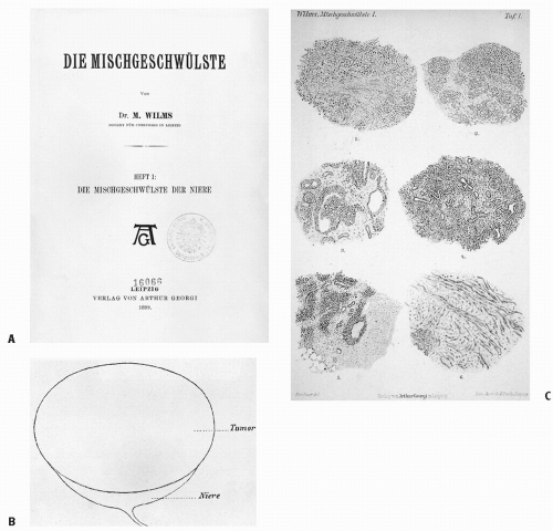
Figure 13.1 A,B: In his monograph, Die Mischgeschwulste der Niere (the mixed tumor of the kidney), published in Leipzig in 1899, Max Wilms described, as his first illustrative case, “Niven Tumor von einem 3 jahrien Mädchen” (“renal tumor of a 3-year-old girl”). C: Wilms described the blastemal, epithelial (tubules), and stromal elements seen on microscopic examination of “mixed” renal tumor.


By the mid-20th century, radiation oncologists had developed a large body of clinical experience with Wilms tumor and began formulating opinions about the role of radiotherapy in the management of the tumor. Ralston Paterson (6), distinguished radiation oncologist of the Christie Hospital and Holt Radium Institute of Manchester, England, wrote in his 1948 textbook:


We feel that the most promising policy’s to start treatment by radiotherapy and not by surgery. X-ray therapy is used and consists of abdominal x-ray baths to cover the entire tumor and any possible intra-abdominal extension. An interval of 6 weeks to 3 months is then allowed in which the tumor mass entirely disappears. … Two opposing fields, anterior and posterior … cover the whole abdomen including the liver. … The essential point in the treatment of Wilms tumor is to remember that even enormous tumors can be made operable and that radiation followed by nephrectomy can undoubtedly obtain cure in some cases.

Dean and Guttmann (7) expressed the predominant viewpoint of US radiation oncologists when they wrote in their 1950 textbook:


The successful treatment of a Wilms tumor depends upon the complete removal or complete devitalization of the primary growth before metastasis occurs. No type of treatment given singly or combined with other forms of treatment has proved successful after metastasis has become established. Ladd, working at the Children’s Hospital of Boston, has obtained by far the best end results by operating on these infants as soon as possible. We also recommend prompt removal … [When preoperative therapy is necessary] … to shrink a Wilms tumor and thereby facilitate its operable removal, external roentgen therapy is the treatment of choice. Usually, however, after five or six exposures, the tumors become significantly smaller and sometimes at the end of 2 weeks it is no longer palpable. … Following radiation infarction seems to be more widespread.

Dean and Guttmann’s views were reinforced by Jacox and Cahill’s (8) review of the management of Wilms tumor. They wrote:


At present we believe that nephrectomy should be performed as soon as the diagnosis of Wilms tumor is made, irrespective of the size of the mass. In general, preoperative irradiation is not favored, because we believe that waiting needlessly jeopardizes the patient’s chances of survival. Ladd and White and their confreres at the Boston Children’s Hospital have made a serious study of the removal of these tumors by the transperitoneal route, with a minimum of handling either for diagnosis or on the operating table, with ligation of the renal vessels and pedicle before displacement of the tumor. The result has been a much higher proportion free from recurrence that has formerly been reported.

What we can see, in this brief historical review of the origins of radiotherapy for Wilms tumor, is the divide between US radiation oncologists and their European colleagues. The European literature favored preoperative treatment, and the US literature favored postoperative radiation therapy. We also see the acknowledgment that wide treatment volumes to cover the entire tumor bed are necessary. We will address these themes in more detail later in this chapter.


EPIDEMIOLOGY

Wilms tumor (nephroblastoma) is an embryonic kidney tumor. It is the most common abdominal tumor in children and represents 6% of childhood cancer. The incidence rate in white children younger than 15 years is 8.1 new cases per million population (9). There are approximately 470-500 new cases in the United States per year (10,11). Wilms tumor is bilateral at presentation in 4-8% of cases (12, 13, 14).

The median age at diagnosis is 41.5 months for boys with unilateral tumors and 46.9 months for girls with unilateral tumors. For bilateral tumors, the median age at presentation is 29.5 months for boys and 32.6 months for girls (11). More than 75% of patients present before 5 years of age. The maleto-female ratio is 0.92 for unilateral tumors and 0.6 for bilateral tumors (11). The incidence rate is approximately three times higher for blacks in the United States and Africa than for East Asians. Rates for the white populations in Europe and North America are intermediate between those of blacks and East Asians. Children tend to present with more advanced disease in less developed nations (11, 12, 13, 14).


MOLECULAR BIOLOGY

In Chapter 5, we discussed the Knudson “two-hit” hypothesis of the origins of retinoblastoma. After vetting his hypothesis for retinoblastoma, Knudson proposed a similar model for Wilms tumor in 1972 (15,16). Like retinoblastoma, Wilms tumor may be unilateral or bilateral. The 4-8% occurrence of bilateral disease, appearing at an earlier age than unilateral disease and associated with a greater frequency of other hereditary anomalies, supports the concept of a specific predisposing constitutional chromosomal deletion. It should be noted that fewer familial and bilateral cases of Wilms tumor than retinoblastoma were available for analysis because of the lower incidence of familial Wilms tumor and the poor disease survival at that time (15, 16, 17).

Wilms tumor is associated with congenital anomalies in 10-13% of cases (16, 17, 18, 19). Aniridia is present in 1% of children with Wilms tumor; hemihypertrophy is noted in 2-3% (19, 20, 21). Other genitourinary malformations are identified in 5% of cases, primarily cryptorchidism, hypospadias, double collecting system, or fused kidney (21,22). It seemed reasonable to look for candidate Wilms tumor genes in association with these congenital anomalies.

The next major clue to the molecular genetics of Wilms tumor was found from the syndrome of Wilms tumor with aniridia, genitourinary malformations, and mental retardation (WAGR syndrome). Karyotypic analysis of children with WAGR syndrome showed a deletion on the short arm of chromosome 11, band 13 (11p13). This deletion encompasses the aniridia gene PA×6 and the Wilms tumor suppressor gene WT1. WT1 is a developmentally regulated transcription factor of the zinc finger family. However, analysis of sporadic Wilms tumors shows evidence of WT1 mutation in only 5-10% of cases. Therefore, although WT1 appears to be a tumor suppressor gene, it accounts for a minority of Wilms tumors (19, 20, 21, 22, 23, 24).

Wilms tumor also occurs with greater frequency in the Beckwith-Wiedemann syndrome (BWS) (25,26). This familial condition variably includes macrosomia or hemihypertrophy, macroglossia, omphalocele, abdominal organomegaly, and ear pits or creases. The genetic locus of this syndrome is also on
the short arm of chromosome 11 (11p15). The putative second Wilms tumor suppressor gene, located at this site, is called WT2. WT2(LP15) has effects on IGF2, the H19 tumor suppressor gene, and the P57 cell cycle regulator (25, 26, 27, 28).

In addition to the tumor suppressor genes associated with Wilms tumor, there is evidence of genetic loci that may be related to more malignant or aggressive Wilms tumors. In a study of patients from National Wilms Tumor Studies (NWTS) 3 and 4, loss of chromosomal material (called loss of heterozygosity [LOH]) on the long arm of chromosome 16 (16q) and short arm of chromosome 1p was associated with inferior outcomes (27,28). In NWTS-5, 2021 children were prospectively evaluated for the poor prognostic significance of tumorspecific LOH for chromosomes 1p or 16q. In clear cell sarcoma of the kidney (CCSK) and rhabdoid tumor (RTK), LOH for 1p and 16q were rarely observed. The results for favorable histology (FH) Wilms tumor are shown in Table 13.1A and B. The incidence of LOH at 1p and 16q was 11.3% and 17.4%, respectively. LOH at either 1p or 16q was only associated with higher risk of relapse for low-stage (stage I/II) patients in comparison to stage III/IV. It was postulated that two-drug chemotherapy was insufficient to overcome the effect of loss of the putative tumor suppressor genes located within these chromosomal regions. Conversely, the more intensive treatment with three drugs did overcome the effect of this loss in stage III/IV patients. The relative risk of relapse and death in low- and high-stage patients was significantly elevated in those patients with LOH at both loci (29). The Children’s Oncology Group (COG) is presently using LOH status at 1p and 16q in addition to tumor stage and histology in stratifying patients according to risk of relapse. Children with low- and high-stage tumors with LOH at both 1p and 16q will have more intensified therapy compared to NWTS-5.

In 2007, a previously unknown gene on the × chromosome WTX was found to be inactivated in approximately one third of tumors. Tumors with mutations in WTX lack WT1 mutations and both genes share a restricted temporal and spatial expression pattern in normal renal precursors. The data suggest that WTX is a Wilms tumor suppressor gene with an important role in normal kidney development. WTX is frequently altered in sporadic tumors by a single somatic event that affects both sexes equally. The one-hit inactivation of a tumor suppressor gene is a departure from the biallelic Knudson model (30). Other known abnormalities in Wilms tumor include activating mutations in the β-catenin gene (CTNNB1) on chromosome 3p22, which often coincide with WT1 mutations (31).


Table 13.1 Analysis for the Joint Effect of LOH at 1p and 16q for (A) Stage I/II and (B) Stage III/IV Favorable Histology Patients (29)


LOH Status


No. of Patients


No. of Relapses


4-Year RFS


RR/p-Value


No. of Deaths


4-Year Overall Survival


RR/p-Value


(A) Stage I/II favorable histology patients


Neither


750


60


91.2


14


98.4


1p only


60


11


80.4


2.19/0.02


4


91.2


4.03/0.02


16q only


114


19


82.5


1.91/0.01


3


98.1


1.40/0.60


Both


46


11


74.9


2.88/0.001


4


90.5


4.25/0.01


(B) Stage III/IV favorable histology patients


Neither


500


82


83.0


38


91.9


1p only


56


6


89.0


0.69/0.37


2


97.6


0.52/0.36


16q only


100


15


85.3


0.89/0.67


7


92.0


0.88/0.76


Both


30


9


65.9


2.41/0.01


5


77.5


2.66/0.04


Another interesting study showed constitutional 11p15 abnormalities in genomic lymphocyte DNA from 3% of patients with nonsyndromic sporadic Wilms tumors, including 12% of bilateral cases. No such abnormalities were detected in control subjects. Such findings may be helpful in assessing the risk of contralateral Wilms tumor development and for targeted surveillance of at-risk family members (32).

In an effort to identify specific biologic markers associated with relapse in FH Wilms tumor, the COG conducted a study to determine the feasibility and potential clinical utility of classifiers of relapse based on global gene expression analysis. There were no significant genetic markers for relapse in stages I and II tumors. However, for stage III tumors the classifiers developed using 50 genes were associated with relapse with a sensitivity of 47% and specificity of 70%. This compares with a sensitivity of 8% and specificity of 96% for LOH of both 1p and 16q in stages III and IV FH Wilms tumor. Analysis of specific genes revealed apoptosis, IGF-1 signaling, Wnt/β-catenin pathway, 1q gain, and epigenetic modification to be mechanisms important in relapse. Two additional potential therapeutic targets identified in this study were FRAP/MTOR and CD40. All these pathways are potential targets for future therapies (33).


PATHOLOGY

The childhood kidney tumor pathology classification systems are those of the NWTS (Table 13.2) and the International Society of Pediatric Oncology (SIOP) study (Table 13.3) and COG (Table 13.4).


Mesoblastic Nephroma

Mesoblastic nephroma is the most common renal tumor encountered in the first month of life. Its median age at presentation is 3 months. It is distinguished from Wilms tumor by its usually benign behavior, a preponderance of mesenchymal derivatives, and a lack of the malignant epithelial components
seen in Wilms. The tumor consists of spindle-shaped cells in interlacing bundles adjacent to renal parenchyma where there are foci of cystic or dysplastic tubules. The treatment of choice is nephrectomy. Local recurrence is unusual. Nonetheless, adequate margins of resection should be obtained, although recurrence even after operative rupture or positive margins at resection is rare. Distant metastases are also rare. The actuarial 2-year survival rate is excellent at 98% (34, 35, 36, 37).


Table 13.2 Classification of Pediatric Renal Tumors


International Society of Pediatric Oncology


National Wilms Tumor Study


I. Low risk


Cystic partially differentiated Wilms tumor


Mesoblastic nephroma


Mesoblastic nephroma


Wilms tumor with fibroadenomatous structures


Highly differentiated epithelial Wilms tumor


II. Intermediate risk


Favorable histology


Nonanaplastic Wilms tumor Wilms tumor with its variants (excluding low-risk types)


Favorable histology Wilms tumor


III. High risk


Anaplastic Wilms tumor


Anaplastic Focal Diffuse


Clear cell sarcoma


Clear cell sarcoma


Rhabdoid tumor


Rhabdoid tumor


Modified from Schmidt D, Beckwith JB. Histopathology of childhood renal tumor. Hematol Oncol Clin North Am. 1995;9:1179-1200, with permission.


Table 13.3 Revised International Society of Pediatric Oncology Working Classification of Renal Tumors of Childhood (2001) for Pretreated Cases


Low-risk tumors


Mesoblastic nephroma


Cystic partially differentiated nephroblastoma


Completely necrotic nephroblastoma


Intermediate-risk tumors


Nephroblastoma, epithelial type


Nephroblastoma, stromal type


Nephroblastoma, mixed type


Nephroblastoma, regressive type


Nephroblastoma, focal anaplasia


High-risk tumors


Nephroblastoma, blastemal type


Nephroblastoma, diffuse anaplasia


Clear cell sarcoma of the kidney


Rhabdoid tumor of the kidney


From Vujanic GM, Sandstedt B, Harms D, et al. Revised International Society of Paediatric Oncology (SIOP) working classification of renal tumors of childhood. Med Pediatr Oncol. 2002;38:79-82, with permission.


Table 13.4 Children’s Oncology Group Classification of Renal Tumors


Nephroblastic tumors


Nephroblastoma Favorable histology Anaplasia (diffuse, focal)


Nephrogenic rests and nephroblastomatosis


Cystic nephroma and cystic partially differentiated nephroblastoma


Metanephric tumors Metanephric adenoma Metanephric adenofibroma Metanephric stromal tumor


Mesoblastic nephroma


Cellular, classic, mixed


Clear cell sarcoma


Rhabdoid tumor


Renal epithelioid tumors of childhood


Angiolipoma


Ossifying renal tumor of infavcy


Nodular Renal Blastemas and Nephrogenic Rests

A spectrum of “pre-Wilms tumor” entities has been described. Nodular renal blastemas are small but visible sub capsular nodules composed of benign embryonic rests. Nephrogenic rests may be limited to the periphery of the renal cortex (perilobar) or randomly distributed throughout the renal lobe (intralobar). Multifocal or diffuse nephrogenic rests are called nephroblastomatosis.


Wilms Tumor

Wilms tumor is a triphasic embryonal neoplasm, which includes blastemal, epithelial (tubules), and stromal elements. Each element may exhibit a variety of patterns of aggregation or lines of differentiation (Fig. 13.2A-C) (35, 36, 37). The proportion of the three components varies from tumor to tumor. If one of the components comprises more than two thirds of the tumor sample, the pattern is designated according to the predominant component. The mixed type is most common (41% of Wilms tumor), followed closely by the clinically more aggressive blastemal predominant (39%), the more indolent epithelial predominant (18%), and the stromal predominant (1%), which behaves like the mixed type (35, 36, 37).

The gross pathologic features of Wilms tumor include its general occurrence as a single unilateral tumor, although multicentric growth and bilateral disease can occur. The tumor is typically solid, lobulated, and not calcified. However, soft and cystic areas may be encountered.


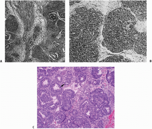
Figure 13.2 A,B: James B. Ewing, renowned for the four editions of his book Pathology of Neoplasia (Philadelphia, PA: WB Saunders; 1942) and his identification for the tumor now called Ewing sarcoma, also provided excellent histologic descriptions of Wilms tumor. These slides, from Ewing’s book, show the topography of Wilms embryonal tumor of the kidney with (A) epithelial tubules lining in masses of spindle and polyhedral cells and (B) the small round cells of the embryonal tumor infiltrating adjacent tissue. C: Favorable histology triphasic Wilms tumor with predominantly epithelial (tubular) differentiation (arrow).

Histopathologic studies in the NWTS identified factors that correlate with prognosis. In the first NWTS, 88% of cases were categorized as FH, defined as having typical histologic features of Wilms tumor without anaplastic or sarcomatous components (38). The frequency of this pattern was upheld in the third NWTS, with 89% of cases categorized as FH on central pathology review (39).

Three entities traditionally have been grouped under the general term unfavorable histology (UH) in the NWTS: anaplastic Wilms tumor, CCSK, and RTK (36). The latter two are no longer considered to be variants of Wilms tumor but are distinct entities (35).

Anaplasia is defined as the significant enlargement of nuclei in the stromal, epithelial, or blastemal cell lines to at least three times the diameter of adjacent nuclei of the same cell type; hyperchromatism of these enlarged nuclei; and multiple mitotic figures. DNA indices greater than 1.5 are associated with anaplastic histology (Fig. 13.3A and B) (35,37,40,41). Anaplasia was noted in 4% of all NWTS-3 entries and in 5% of patients in the SIOP studies (35,37,40,41). Anaplastic tumors are extremely rare in infants, are uncommon before 2 years of age, and make up about 10% of Wilms tumors diagnosed after 5 years of age (37). Anaplasia appears to be associated with greater resistance to chemotherapy rather than greater aggressiveness of Wilms tumor. The 4-year survival rate in NWTS-3 for patients with anaplastic histology was 82% but was lower in earlier studies (38, 39, 40, 41).

Anaplastic Wilms tumor may be focal or diffuse. When this distinction was originally drawn, the term diffuse anaplasia was applied to tumors with anaplastic nuclear changes in more than 10% of 400 microscopic fields. Focal anaplasia was applied to the remainder of anaplastic tumors. Using this distinction, focal anaplasia was associated with a more favorable outcome than diffuse anaplasia in the first NWTS, but this difference did not obtain statistical significance and was not confirmed in the second and third NWTS trials (42).

The original distinction between focal and diffuse anaplasia did not consider distribution of tumor throughout
the kidney, which might affect the likelihood of complete resection. In a recently revised definition, focal anaplasia refers to anaplasia that is sharply localized in the primary tumor, without significant nuclear or mitotic atypia in the remainder of the lesion. Diffuse anaplasia is either nonlocalized anaplasia, localized anaplasia with severe nuclear unrest elsewhere in the tumor, anaplasia outside the tumor capsule or in metastases, or anaplasia found in a random biopsy taken from the tumor.


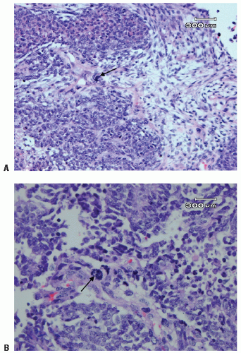
Figure 13.3 Anaplastic Wilms tumor with a large dark hyperchromatic nucleus (arrow) (A) and multipolar mitosis (arrow) (B).

Using the new criteria, and on review of patients in NWTS-3 and NWTS-4, patients with focal anaplasia had a 4-year survival rate statistically significantly higher than that of patients with diffuse anaplasia (97% vs. 50%). This difference did not exist for stage I tumors (100% survival for focal or diffuse) but did exist for stages II (90% vs. 55%), III (100% vs. 45%), and IV (100% vs. 4%) (37,42).

Patients with anaplastic histology Wilms tumor were treated in prospective single-arm studies in NWTS-5. Patients with stage I anaplastic tumors were treated with vincristine and dactinomycin for 18 weeks without radiation therapy. Patients with stages II-IV diffuse anaplastic histology were treated with regimen “I” chemotherapy that consisted of vincristine, doxorubicin, cyclophosphamide, and etoposide for 24 weeks plus flank/abdominal radiation therapy. Among 2596 patients with Wilms tumor enrolled onto NWTS-5, 281 (10.8%) had anaplastic histology. The 4-year event-free and overall survival rates for stage I anaplastic histology tumors were 70% and 83%, respectively. The 4-year event-free and overall survival rates in stages II, III, and IV diffuse anaplastic tumors after immediate nephrectomy were 83% and 82%, 65% and 67%, and 33% and 33%, respectively. These results form the basis for the COG study with augmentation of therapy for stages I, III, and IV anaplastic tumors (43).


Rhabdoid Tumor of the Kidney

RTK is a highly malignant tumor characterized by uniform cellular infiltrates initially interpreted as rhabdomyoblastic or sarcomatous elements. The tumor is unrelated to rhabdomyosarcoma or Wilms tumor and may be of neural crest origin (35, 36, 37). Rhabdoid cells are characterized by eosinophilic cytoplasm that contains hyaline globular inclusions. On electron microscopy, these inclusions are found to be intermediate filaments; most contain vimentin and cytokeratin. The nuclei are large, round, and vesicular, often containing a centrally placed eosinophilic nucleolus (Fig. 13.4) (37).

Most RTK of the kidney are diagnosed in the first 2 years of life. RTK have also been reported as primary extrarenal lesions; there is a known association between RTK and primary central nervous system neoplasms (37). Children with RTK have responded poorly to the therapies of the NWTS. The 4-year relapse-free survival rate for patients treated with vincristine, actinomycin D, and adriamycin on NWTS-3 was 23%, and the 4-year overall survival rate was 25% (39,44).


Clear Cell Sarcoma of the Kidney

CCSK is a primitive mesenchymal neoplasm that makes up 4% of childhood renal tumors. The cell of origin is unknown. The lesion is distinguished by cells with poorly stained cytoplasm. The cell boundaries are indistinct, and cytoplasmic vacuolation may be prominent. The classic histologic pattern has a characteristic arborizing network of thin-walled capillary blood vessels that separates groups of cells (Fig. 13.5A and B) (37,45). In NWTS-3, 23% of children with CCSK developed bone metastases, compared with 0.3% of all other
children entered in the study. Response of CCSK to therapy was poor until adriamycin and local irradiation were added to the treatment program. The 4-year relapse-free survival rate for patients with stages I-IV CCSK treated with vincristine, adriamycin, and actinomycin D on NWTS-3 was 71% (39).


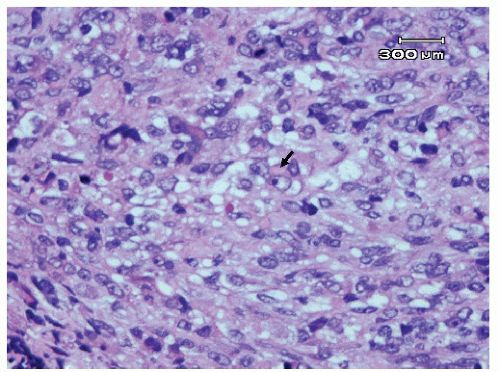
Figure 13.4 Rhabdoid tumor (RTK) with tumor cells with large eccentric vesicular nuclei, prominent nucleoli, and cytoplasmic inclusions (arrow).


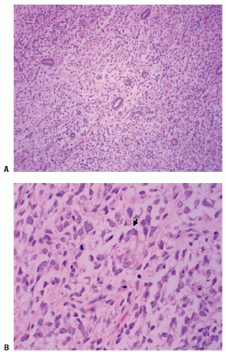
Figure 13.5 Clear cell sarcoma (CCSK) with tumor cells composed of uniform clear cytoplasm surrounding entrapped renal tubules: (A) low power, (B) high power showing prominent vacuolated cytoplasm (arrow).

A total of 86 patients with CCSK were registered in NWTS-4. This study was designed to compare the efficacy and toxicity of a new schedule of administration of dactinomycin and doxorubicin. There was no significant difference between those patients initially randomized to standard and pulse-intensive (PI) chemotherapy with vincristine, dactinomycin, and doxorubicin. The 8-year relapse-free and overall survival rates were 72% and 87% for PI and 70% and 84% for standard chemotherapy, respectively. The second randomization was to short-duration (no further chemotherapy) and long-duration (additional 9 months) chemotherapy. Again, there was no significant difference in survival between the two arms. The 8-year relapse-free and overall survival rates were 61% and 86% for the short-duration chemotherapy and Even though the relapse-free survival rates were not significantly different (p = 0.08) the long-duration arm resulted in a higher relapse-free survival rate. The overall survival rate in NWTS-4 (83%) was significantly higher than in NWTS-3 (67%) (46).


CLINICAL PRESENTATION AND WORKUP

As we have already noted, several clinical syndromes are risk factors for the development of Wilms tumor. These include aniridia, BWS (exomphalos, macroglossia, and gigantism), and hemihypertropia. If somatic changes of these kinds are known to exist, then routine screening in an attempt to make the early diagnosis of Wilms tumor is appropriate (47). A physical examination and periodic ultrasound are indicated. It is interesting to note that in Germany, 10% of patients with Wilms tumor are diagnosed at infant and childhood screening examinations, conducted for known predisposing syndromes, and at routine well-baby examinations (47).

The majority of children with Wilms tumor are diagnosed in response to a medical complaint causing a visit to the doctor; the clinical presentation usually is an abdominal mass (83%), fever (23%), or hematuria (21%). Abdominal pain (37%) may be the result of local distention, spontaneous intralesional hemorrhage, or peritoneal rupture. In one major series, abdominal pain correlated with poorer 5-year survival (48). Less common presenting signs and symptoms include hypertension, varicocele, hernia, enlarged testicle, congestive heart failure, hypoglycemia, Cushing syndrome, hydrocephalus, pleural effusion, and an acute abdomen (49).

The presence and character of the abdominal pain, the previous medical history, and the family history are important aspects of the medical interview. Physical examination is of value in assessing abdominal status and identifying associated congenital anomalies.

The optimum diagnostic imaging workup for Wilms tumor is a matter of controversy. Evaluation of disease extent classically included intravenous pyelogram (IVP) and chest radiograph. Current imaging protocols largely replace the IVP with abdominal ultrasound. Ultrasound usually allows determination of the origin of a childhood abdominal mass, identifies a contralateral kidney, and demonstrates the presence or absence of tumor extension into the renal vein or inferior vena cava (50).

When an abdominal mass is identified or suspected, abdominal computed tomography (CT) is used to assess the volume of tumor involvement in one or both kidneys, renal function, retroperitoneal lymph nodes, and invasion of the collecting system or renal vein; evaluate the margin between tumor, kidney, and surrounding structures; assess hepatic metastasis (although many children thought to have invasion of the liver from a right-sided Wilms tumor are found at surgery to have hepatic compression rather than invasion); and demonstrate lesions in the opposite kidney, which may represent either bilateral Wilms tumor or nephrogenic rests (Fig. 13.6) (50). The evaluation of the contralateral kidney has become a matter of discussion in the surgical literature. Some authorities believe that preoperative CT and magnetic resonance imaging (MRI) can be used to rule in or rule out
bilateral Wilms tumor. Others believe that imaging is useful but not definitive and that a surgical exploration of the contralateral kidney remains essential during the surgical approach to the primary tumor (17,47).


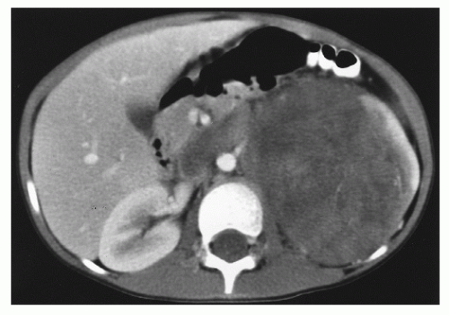
Figure 13.6 A 5-year-old girl presented with intermittent sharp abdominal pain, nausea, vomiting, weight loss, and hematuria. Abdominal computed tomography showed a 7.9- to 12.5-cm mass at the left renal region, beginning immediately subdiaphragmatically and extending to just below the aortic bifurcation. There was a calcified rim superiorly and a heterogeneous parenchymal enhancement. Left renal vein invasion and inferior vena cava thrombosis were observed. On exploratory laparotomy, tumor was palpated in the inferior vena cava. The tumor was resected, with some tumor spillage from a weak point on Gerota fascia. Tumor thrombus was removed from the inferior vena cava. Pathology showed a favorable histology Wilms tumor, 13.5 cm in greatest diameter, invading through the renal capsule to the inked surgical margin. Rupture of the renal capsule was identified. Tumor was present at the renal vein margin of the tumor thrombosis with focal invasion of the vein wall. No lymph nodes were involved. For pathologic stage III Wilms tumor, favorable histology, the child was treated with chemotherapy and flank irradiation. There was no evidence of persistent or recurrent tumor 3 years after diagnosis.

Plain chest radiographs should be obtained to determine whether pulmonary metastases are present. Some centers recommend thoracic CT to detect pulmonary metastasis that might be missed on chest radiograph. CT-positive, chest radiograph-negative lung metastases do occur (50). There has been much uncertainty regarding appropriate therapy for pulmonary disease documented only by CT in the presence of normal plain chest films (51). Radionuclide bone scan is indicated in CCSK and renal RTK. Bone scan and skeletal survey are complementary in CCSK. If only one technique is used, metastases may be missed (38,52). Cranial CT is of value in RTK and, perhaps, in Wilms tumor with overt pulmonary involvement at diagnosis.


STAGING

A staging system for Wilms tumor was first published by Cassady et al. (53,54) in 1973, building on prognostic factors identified in a review from Garcia et al. (55). The Cassady system remains useful for determining whether young children with early stage disease may be appropriate for therapy with surgery alone. For tumor confined to the kidney and completely excised in children younger than 2 years of age at diagnosis and with tumors that weigh less than 550 g, the prognosis is excellent. Such patients may be treated appropriately with surgery alone and are considered to have stage I disease (54, 55, 56). The absence of an inflammatory pseudocapsule, renal sinus invasion, capsular invasion, and intrarenal vessel invasion in such patients are additional factors that mitigate against the risk of relapse when treated with nephrectomy alone (50).

The NWTS proposed initial staging criteria to prospectively address tumor-related factors. The first two NWTS studies were based on a system of surgically identified groups (38).

Beginning with the third NWTS trial in 1979, a new staging system was adopted. Specific disease-related parameters noted to be prognostically significant in analyses of NWTS-1 and NWTS-2 led to modifications of the system and the change from groups to stages. A closed or open biopsy permitted categorization as stage II (rather than group III), assuming subsequent total tumor removal. Local spill of tumor during surgery was downstaged from group III to stage II, reflecting data indicating that such cases had excellent tumor control even with limited irradiation and chemotherapy in NWTS-1 and NWTS-2 (57). Also, the NWTS-3 staging system upstages all previous group II cases with lymph node metastasis to stage III, even when all visible disease has been completely resected. The adverse impact of lymph node involvement, both on abdominal recurrence and on ultimate relapse-free survival, has been documented (13,22,38,58).

For NWTS-5, the most significant change was the distinction between stages I and II. Criteria for stage I was refined to accommodate an important subset of patients who were being managed by nephrectomy alone. Before NWTS-5, the distinction between stages I and II in the renal sinus was established by the hilar plane, which was an imaginary plane connecting the most medial aspects of the upper and lower poles of the kidney. This criterion was difficult to apply because of tumor distortion, and thus the hilar plane criterion has been replaced with renal sinus vascular or lymphatic invasion. This definition includes not only the involvement of vessels within the hilar soft tissue, but of vessels located in the radial extensions of the renal sinus into the renal parenchyma (59,60).

The COG staging guidelines for Wilms tumor are shown in Table 13.5. These guidelines are essentially similar to NWTS-5 except for the fact that children with tumor spillage are upstaged from stage II to stage III because of the higher risk for relapse with two-drug chemotherapy alone (61).

In the SIOP Wilms tumor trial 1, investigators compared preoperative radiotherapy with primary surgery. The staging system used in this trial is shown in Table 13.6. SIOP subsequently began using NWTS grouping and staging, with the proviso that the NWTS systems were not specifically designed for the preoperative therapy often used by SIOP (62, 63, 64, 65, 66, 67, 68, 69, 70, 71). For this reason, outcomes between NWTS and SIOP studies should not be compared stage-for-stage. In addition, SIOP uses a “stage II, node negative” and “stage II, node positive” distinction and therefore includes some NWTS stage III tumors in the stage II infradiaphragmatic SIOP stage.


Table 13.5 Children’s Oncology Group Staging of Wilms Tumor


Stage I


Tumor limited to kidney, completely resected. The renal capsule is intact. The tumor was not ruptured or biopsied prior to removal. The vessels of the renal sinus are not involved. There is no evidence of tumor at or beyond the margins of resection.
Note: For a tumor to qualify for certain therapeutic protocols as Stage I, regional lymph nodes must be examined microscopically.


Stage II


The tumor is completely resected and there is no evidence of tumor at or beyond the margins of resection. The tumor extends beyond kidney, as is evidenced by any one of the following criteria:
There is regional extension of the tumor (i.e., penetration of the renal capsule, or extensive invasion of thesoft tissue of the renal sinus, as discussed below).
Blood vessels within the nephrectomy specimen outside the renal parenchyma, including those of the renal sinus, contain tumor.
Note: Rupture or spillage confined to the flank, including biopsy of the tumor, is no longer included in Stage II and is now included in Stage III.


Stage III


Residual nonhematogenous tumor present following surgery, and confined to abdomen. Any one of the following may occur:
Lymph nodes within the abdomen or pelvis are involved by tumor. (Lymph node involvement in the thorax or other extra-abdominal sites is a criterion for Stage IV.)
The tumor has penetrated through the peritoneal surface.
Tumor implants are found on the peritoneal surface.
Gross or microscopic tumor remains postoperatively (e.g., tumor cells are found at the margin of surgical resection on microscopic examination).
The tumor is not completely resectable because of local infiltration into vital structures.
Tumor spillage occurring either before or during surgery.
The tumor was biopsied (whether tru-cut, open, or fine needle aspiration) before removal.
Tumor is removed in greater than one piece (e.g., tumor cells are found in a separately excised adrenal gland; a tumor thrombus within the renal vein is removed separately from the nephrectomy specimen).


Stage IV


Hematogenous metastases (lung, liver, bone, brain, etc.), or lymph node metastases outside the abdominopelvic region are present. (The presence of tumor within the adrenal gland is not interpreted as metastasis and staging depends on all other staging parameters present.)


Stage V


Bilateral renal involvement by tumor is present at diagnosis. An attempt should be made to stage each side according to the above criteria on the basis of the extent of disease.


NWTS, SIOP, AND THE MANAGEMENT OF WILMS TUMOR

The NWTS, SIOP, and the United Kingdom Children’s Cancer Study Group (UKCCSG) have compiled a large body of information concerning the clinical management of Wilms tumor. The most widely quoted studies are those of NWTS and SIOP.


The NWTS and SIOP Strategies

The SIOP studies began with the presumption that treatment with radiation therapy or chemotherapy before surgery would render a Wilms tumor less vulnerable to intraoperative rupture and surgery-related tumor seeding. Downstaging the tumor was also hoped to reduce treatment-related morbidity by reducing the total amount of treatment (62, 63, 64). In accordance with this philosophy, the initial strategy of SIOP was to evaluate the roles of radiotherapy and chemotherapy before definitive surgery based on clinical diagnosis. Subsequently, treatment was assigned according to the extent of disease found at surgery. One disadvantage is the risk of misdiagnosis and mistreatment (i.e., treating a tumor other than Wilms tumor or treating benign disease). In SIOP 9, the error rate (i.e., the proportion of patients who were found not to have Wilms tumor) was 6%. In addition, the SIOP approach runs the risk of obscuring important prognostic clues in individual patients (64). Lymph node involvement and histologic subtype may be affected by preoperative therapy. A particular drawback of preoperative therapy is that one may miss diffuse anaplasia. Because prognosis and subsequent therapy are influenced by histology and lymph node involvement, the SIOP approach may render therapy less precise by interfering with histology obtained at the time of definitive surgery. Supporting this contention was a review of data from two SIOP studies indicating that necrotic changes were more frequently seen in patients pretreated with radiotherapy than in those pretreated with chemotherapy. However, an analysis of
posttreatment histology in these two SIOP studies and in 83 patients who underwent prenephrectomy chemotherapy in NWTS-3 showed no evidence that prenephrectomy therapy altered the detection of anaplastic histology (62, 63, 64). It appears that the SIOP presurgical therapy approach does not influence histology.


Table 13.6 Staging System Used in SIOP Wilms Tumor Trial 1


Stage I


The tumor is limited to the kidney and is completely excised


Stage II


The tumor extends outside the kidney but is completely excised


Stage III


Incomplete excision of the tumor but without hematogenous metastatic spread


Stage IV


Hematogenous metastases are present


Stage V


Bilateral renal tumors


The NWTS strategy is to forgo preoperative therapy in order to obtain the maximum amount of information concerning prognostic factors and tailor therapy accordingly. Therefore, the local extent of the primary tumor, degree of anaplasia, presence of unusual histology, and presence or absence of lymph node involvement are assessed in the absence of the confounding influence of preoperative chemotherapy or radiotherapy in order to select therapy. The benefits of the NWTS approach are the generation of a large body of information about prognostic clues, the avoidance of misdiagnosis, and the customization of therapy.


The National Wilms Tumor Studies

NWTS-1 (1969-1974) asked several important treatment questions: Is postoperative radiotherapy necessary in group I disease? Is single-agent chemotherapy with either vincristine or actinomycin D equivalent to combining these drugs for group II and III disease? Is preoperative vincristine of value in group IV disease? The study is shown schematically in Figure 13.7, and the results are summarized in Table 13.7 (38). Radiotherapy dosages were age adjusted (birth to 18 months of age, 18-24 Gy; 18-30 months, 24-30 Gy; 31-40 months, 30-35 Gy; 41 months or older, 35-40 Gy). Radiotherapy appeared to be unnecessary in group I babies; combined drug therapy was superior in groups II and III; and preoperative vincristine was not helpful in group IV. Patients older than 2 years with group I tumors showed an advantage with irradiation. However, patients with group II tumors receiving actinomycin D with vincristine seemed to do as well as those with group I disease. It was postulated that postoperative vincristine with actinomycin D could substitute for postoperative radiotherapy with actinomycin D (38,72). This study demonstrated the significance of the UH versus FH distinction, with a 2-year relapse-free survival rate of 29% for UH and 89% for FH (72,73). Large tumor size, lymph node involvement, and age older than 2 years were confirmed as poor prognostic factors. No radiation dose-response relationship
was discerned in the 10- to 40-Gy range, delays of up to 10 days in initiating postoperative irradiation appeared acceptable, and whole- abdomen irradiation was not found to be necessary for tumor spills confined to the flank or for prior tumor biopsy (17,41,57). In these patients, limited radio therapy fields sufficed.


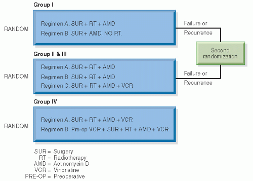
Figure 13.7 The design of NWTS-1. (From D’Angio GJ. National Wilms’ Tumor Study. Seattle, WA: NWTS Data and Statistical Center; 1991 [Informational Bulletin #19], with permission.)


Table 13.7 NWTS-1 Results


Group and Therapy


n


4-Year Relapse-Free Survival (%)


4-Year Survival (%)


Stage I. Age <2 years


38


89


94


A.


Radiotherapy


38


89


94


B.


No radiotherapy


41


88


90


Stage I. Age > 2 years


A.


Radiotherapy


42


76


98


B.


No radiotherapy


42


57


81


Stages II. and III.


A.


AMD


63


56*


71*


B.


VCR


44


57*


71*


C.


AMD and VCR


63


79


84


Stage IV.


A.


Immediate surgery


13


83*a


B.


Preoperative VCR


13


29*a


* p < 0.02.a 2-year survival.


AMD, actinomycin D; VCR, vincristine.


From Refs. 38,57,72, and 75 with permission.


NWTS-2 (1974-1979) explored three major questions: Can vincristine and actinomycin D substitute for radiotherapy in older children with group I disease? Are adjuvant vincristine and actinomycin D for protracted periods helpful in group I? Is the addition of adriamycin to actinomycin D and vincristine of value in groups II to IV? The study is shown schematically in Figure 13.8 (74). Flank irradiation was given in groups II-IV disease according to the same age-dependent scale as NWTS-1. Whole-abdomen irradiation was reserved for diffuse peritoneal seeding. Lung metastases were initially treated with 14 Gy of whole-lung irradiation (WLI), but when a 10% incidence of pneumonitis occurred, the dosage was scaled back to 12 Gy. Study results are summarized in Table 13.8. About 85% of the patients were FH and 15% UH. Two-year survival rates were 54% for UH, 90% for FH, 54% for lymph node positive, and 82% for lymph node negative. Excellent survival rates appeared to be achievable without irradiation in group I, and adriamycin added considerable benefit in groups II-III FH and some benefit in groups II-III UH and group IV. The 2-year relapse-free survival rate for all NWTS-2 patients was 88% for group I, 78% for group II, 70% for group III, and 49% for group IV (17,74, 75, 76).

NWTS-3 (1979-1985) incorporated two major changes in treatment planning: Patients were stratified by stages rather than by groups and the distinction between FH and UH was incorporated into the treatment algorithm (Fig. 13.9). Although data from NWTS-1 and NWTS-2 were analyzed by histology, histology was not used to stratify treatment (38,50,74).


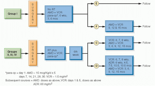
Figure 13.8 The design of NWTS-2. (From D’Angio GJ, Tefft M, Breslow N, et al. Radiation therapy of Wilms’ tumor: results according to dose, field, post-operative timing and histology. Int J Radiat Oncol Biol Phys. 1978;4:769-780, with permission.)


Table 13.8 NWTS-2 Results


Group and Therapy


n


3-Year Relapse-Free Survival (%)


3-Year Survival (%)


Stage I


E. 6 months, ADR, VCR


88


89


97


F. 15 months, ADR, VCR


91


84


92


Stages II and III FH


C.


VCR, AMD


121


70*


82


D.


VCR, AMD, ADR


11


88


92


Stages II and III UH


C.


VCR, AMD


16


35


38


D.


VCR, AMD, ADR


19


42


78


Stage IV


C.


VCR, AMD


22


43*


44


D.


VCR, AMD, ADR


27


60*


60


Lymph node status


Negative or not examined


383


82a


Positive


84


54


* p < 0.05.a 2-year survival.


ADR, adriamycin; VCR, vincristine; FH, favorable histology; AMD, actinomycin D; UH, unfavorable histology.


From Refs. 74 and 75 with permission.


NWTS-3 considered five major questions: Can the duration of chemotherapy be shortened for stage I FH? Can radiotherapy be eliminated for stage II FH? What is the minimum effective radiotherapy dosage for stage III FH? Is cardiotoxic
adriamycin clearly beneficial and therefore necessary in stages II and III FH? Will the addition of cyclophosphamide improve survival in stages I-III UH and in stage IV FH and UH?


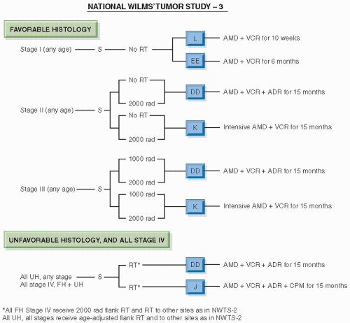
Figure 13.9 The design of NWTS-3. (From D’Angio GJ, Evans A, Breslow N, et al. The treatment of Wilms’ tumor: results of the Second National Wilms’ Tumor Study. Cancer. 1981;47:2302-2311, with permission.)

NWTS-3 results are summarized in Tables 13.9 and 13.10. Short-course therapy appeared equivalent to longcourse therapy in stage I FH. Patients in this group can be treated successfully with a 10-week program of vincristine and actinomycin D without irradiation and can achieve a 4-year relapse-free survival rate of 89% and overall survival rate of 96%. Elimination of radiotherapy was acceptable in stage II FH. The 4-year relapse-free and overall survival rates for the patients not receiving irradiation were 87% and 91%, respectively. In stage III FH, 10 Gy was equivalent to 20 Gy. There was no statistically significant difference in frequency of intra-abdominal relapse between 10 and 20 Gy, although the trend favored the use of adriamycin or irradiation (intr-aabdominal relapse for vincristine, actinomycin D, and 10 Gy, 7 out of 61, or 11%; vincristine, actinomycin D, and 20 Gy, 3 out of 68, or 4%; vincristine, actinomycin D, adriamycin, and 10 Gy, 3 out of 70, or 4%). In an analysis of patients with stage III, local relapse with doxorubicin occurred in 4 out of 134 patients, whereas without doxorubicin, it occurred in 11 out of 141 patients, suggesting that chemotherapy played a role in establishing local control. The addition of adriamycin was not clearly beneficial in stage II but seemed to be of benefit in stage III FH. Cyclophosphamide did not benefit stage IV FH. The 4-year survival rate for stage IV FH treated with vincristine, actinomycin D, adriamycin, and abdominal and lung irradiation was 81%. The use of cyclophosphamide seemed to help patients with focal anaplasia. Patients with CCSK did well, whereas those with RTK continued to do poorly (17,22,39,50).

NWTS-4 (1986-1994) addressed issues of minimization of therapy (and, presumably, therapy-related toxicity) and customization of therapy by stage and histology. The trial was the first study of a pediatric population to evaluate the economic impact of two different treatment approaches, one of which entailed fewer clinic visits. By the end of NWTS-3 it had been shown that 62% of patients with Wilms tumor (stages I-II, FH) needed neither irradiation nor adriamycin. The study was designed to compare the relapse-free and overall survival rates of patients with stages I and II FH and stage I anaplastic tumors treated with conventional actinomycin D and vincristine and with pulsed, intensive actinomycin D and vincristine. Stages III and IV FH and stages I-IV CCSK were treated with conventional actinomycin D, vincristine, and
adriamycin, compared with pulsed, intensive actinomycin D, vincristine, and adriamycin; all patients received radiotherapy (for FH, 10.8 Gy to the abdomen; 12 Gy WLI where appropriate; for stages II-IV anaplastic tumors, a sliding scale of radiation dosage was used, as in NWTS-1). Stages II-IV anaplastic Wilms tumors were treated with appropriate irradiation followed by actinomycin D, vincristine, and adriamycin, compared with these three drugs plus cyclophosphamide. Stages II-IV FH and stages I-IV CCSK were treated with 26 or 54 weeks of chemotherapy (Fig. 13.10).


Table 13.9 NWTS-3 Results


Group and Therapy


n


4-Year Relapse-Free Survival (%)


4-Year Survival (%)


Stage I FH


L. 10 weeks ADR, VCR


306


89


96


EE. 6 months ADR, VCR


301


92


97


Stage II FH


DD. No radiotherapy; AMD, VCR, ADR


70


88


94


DD2. 20 Gy; AMD, VCR, ADR


71


87


90


K. No radiotherapy; AMD, VCR


67


87


91


K2. 20 Gy; AMD, VCR


70


90


95


Stage III FH


DD1. 10 Gy; AMD, VCR, ADR


68


82


91


DD2. 20 Gy; AMD, VCR, ADR


66


86


87


K1. 10 Gy; AMD, VCR


71


71


85


K2. 20 Gy; AMD, VCR


70


77


85


Stage IV FHa


DD. AMD, VCR, ADR


64


72


78


J. AMD, VCR, ADR, CPM


56


78


87


Stages I-III UHa


DD. AMD, VCR, ADR


69


67


68


J. AMD, VCR, ADR, CPM


61


62


68


Stage IV UHa


DD. AMD, VCR, ADR


12


58


58


J. AMD, VCR, ADR, CPM


17


53


52


Anaplastica


DD. AMD, VCR, ADR


31


1:80


II-IV


37


1:78


II-IV


38*


J. AMD, VCR, ADR, CPM


17


1:100


II-IV


83


1:100


II-IV


82*


CCSKa


DD. AMD, VCR, ADR


25


71


75


J. AMD, VCR, ADR, CPM


25


60


76


Rhabdoida


DD. AMD, VCR, ADR


13


23


25


J. AMD, VCR, ADR, CPM


18


27


26


* p < 0.05.a Radiotherapy also given; not a randomized radiotherapy question.


FH, favorable histology; ADR, adriamycin; VCR, vincristine; AMD, actinomycin D; CPM, cyclophosphamide; UH, unfavorable histology.


From Refs. 39,44,76 with permission.


Table 13.10 NWTS-3 Relapse Rates


Stage and Dosage


Flank Relapse (8 years) (%)


Beyond Flank (%)


0 Gy


3.2


<15 Gy


1.6


>15 Gy


0


Stage II: 0 Gy


12.8


4.6


Stage II: 20 Gy


0


4


Stages III-IV


10-20 Gy


3.5-11.8


10 Gy


0-1.8


20 Gy


0


From Refs. 39,44,76.


The results of NWTS-4 are shown in Table 13.11. There was no difference in the low-risk or high-risk patients for standard or PI therapy. When one breaks down the results based on histology, stage, standard or PI therapy, and, for the more advanced patients, short or long course of therapy, there are, once again, no significant differences between the various
treatment groups (77,78). Comparing the results from NWTS-4 with those of the UKCCSG Wilms Tumor Study 2, reported contemporaneously, we see surprisingly similar results (Table 13.12) (79). The risk of local recurrence of patients enrolled in NWTS-4 is shown in Table 13.13.


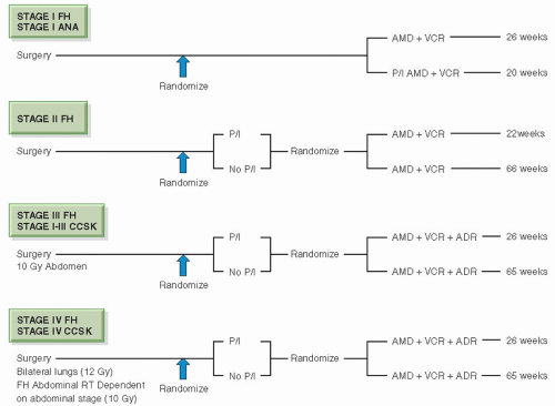
Figure 13.10 NWTS-4 simplified schema. Stage IV anaplastic tumors continued the randomization of NWTS-3. (From Thomas PRM. Wilms’ tumor: changing role of radiation therapy. Semin Radiat Oncol. 1997;7:204-211, with permission.)

The NWTS evaluated the frequency with which spilled tumor cells of FH produce intra-abdominal disease recurrence. Flank irradiation but not doxorubicin reduced abdominal relapse rates. The odds ratio for the risk of recurrence relative to no radiation was 0.35 for 10 Gy and 0.08 for 20 Gy. Tumor spillage resulted in higher relapse and significantly lower survival among stage II patients. For stage II patients (NWTS-4), the 8-year event rates, with and without spillage, respectively, were 79% and 87% for relapse-free survival (p = 0.07), and 90% and 95% for overall survival (p = 0.04) (61).

The evidence from NWTS-4 is that PI initial chemotherapy is as effective as a longer program. Shorter-course subsequent chemotherapy appears as effective as a longer program. Overall survival rates are excellent, and few patients are irradiated (77,78).

The long-term data from NWTS-3 and NWTS-4 show no significant effects for doxorubicin in stage II FH patients; however, for stage III patients, the relapse-free and overall survival rates were 84% and 89% with and 74% and 83% without doxorubicin, respectively. For stage III patients, the use of doxorubicin results in a significant reduction in local and general recurrence without any improvement in overall survival (80).

NWTS-5 (1995-2001) treated stage I favorable and anaplastic histology and stage II FH with 18 weeks of actinomycin D and vincristine. Stage I FH in children younger than 24 months of age and with tumor weight less than 550 g was treated with surgery alone. Stages III-IV FH and stages II-IV focal anaplastic tumors were treated with 24 weeks of actinomycin D, vincristine, adriamycin, and local irradiation and WLI as appropriate. Stages II-IV diffuse anaplastic sarcoma and stages I-IV CCSK were treated with cyclophosphamide, vincristine, adriamycin, and etoposide along with local irradiation and WLI as appropriate. Stages I-IV RTK was treated with carboplatin, etoposide, cyclophosphamide, and radiotherapy. The radiotherapy guidelines in NWTS-5 were similar to those used in NWTS-4 except for anaplastic tumors where a dose of 10.8 Gy was recommended as compared to an age-adjusted schedule used in NWTS 1-4. As a major objective, NWTS-5 sought to evaluate the importance of LOH for chromosome 1p and 16q markers for prognosis. These results have been detailed earlier in the chapter (Table 13.1) (29).

The outcome of 57 patients with FH tumors and peritoneal implants at the time of nephrectomy in NWTS-4 and NWTS-5 were analyzed. Stage III tumors were seen in 74%, and 26% had stage IV tumors. All children received multimodality therapy with three-drug chemotherapy and irradiation. Forty-seven patients (82%) received wholeabdominal radiation therapy and the radiation dose was
10.5 Gy in 50 patients. The overall abdominal and systemic tumor control rates were 97% and 93%, respectively. The detection of peritoneal implants was not associated with inferior survival after this therapy. The 5-year relapse-free survival rates with and without peritoneal implants were 90% and 83%, respectively (81).


Table 13.11 NWTS-4 Results


4-Year Relapse-Free Survival (%)


Low Risk


High Risk


STD


PI


STD


PI


88


84


89


90


Stage and Treatment


Relapse-Free Survival


Stage I


FH-STD


93


FH-PI


95


Ana-STD


93


Ana-PI


88


Stage II


FH-STD + Short


82


FH-STD + Long


88


FH-PI + Short


86


FH-PI + Long


89


Stage III


FH-STD + Short


94


FH-STD + Long


91


FH-PI + Short


92


FH-PI + Long


90


Stage IV


FH-STD + Short


82


FH-STD + Long


93


FH-PI + Short


83


FH-PI + Long


78


STD, standard; PI, pulse intense; FH, favorable histology; ANA, anaplastic; Short, short therapy course; Long, long therapy course;. From Refs. 77 and 78.

Only gold members can continue reading. Log In or Register to continue

Stay updated, free articles. Join our Telegram channel

Jun 19, 2016 | Posted by in GENERAL RADIOLOGY | Comments Off on Wilms Tumor

Full access? Get Clinical Tree

Get Clinical Tree app for offline access