Late Effects of Cancer Treatment


Late Effects of Cancer Treatment


Debra L. Friedman

Louis S. Constine


With advances in multimodality therapy and supportive care, the cure rate for childhood cancer continues to improve and now approaches 80% (1). However, the therapy associated with this improved survival, chemotherapy and radiotherapy (RT), can lead to adverse long-term health-related outcomes that manifest months to years after completion of cancer treatment, commonly called late effects. Late effects include organ dysfunction, subsequent malignant and benign neoplasms, and adverse psychosocial sequelae, placing survivors at risk for chronic health conditions as they enter their adult years. In a report from the Childhood Cancer Survivor Study (CCSS), the 30-year cumulative incidence for severe, disabling, or life-threatening conditions or death due to a chronic condition was 42% (2), and in another report from this same cohort study, the 30-year cumulative mortality was 18% among survivors compared to the general population, and RT increased risk 2.2-fold (3). From the CCSS, where survivors all now have over 20 years of follow-up since childhood cancer diagnosis, Figure 19.1 shows the cumulative incidence of chronic health conditions. Figure 19.2
shows the cause-specific cumulative late mortality. Risk of death remains highest for recurrence of original disease, but 14% of deaths from this cohort are now from subsequent malignancies (4).


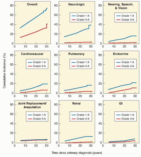
Figure 19.1 Cumulative incidence of chronic health conditions of adult survivors of childhood cancer by primary childhood cancer diagnosis and severity of condition (From Oeffinger KC, Mertens AC, Sklar CA, et al. Chronic health conditions in adult survivors of childhood cancer. N Engl J Med. 2006;355:1572-1582, with permission.)


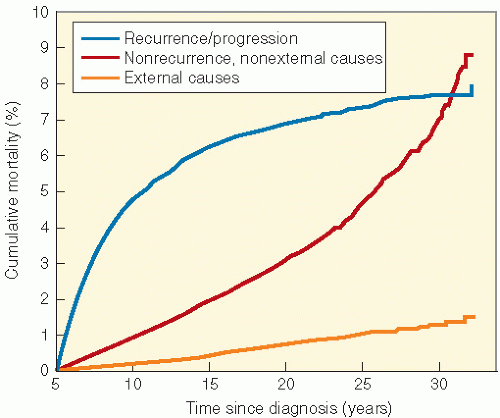
Figure 19.2 Cumulative cause-specific mortality. (From Armstrong GT, Liu Q, Yasui Y, et al. Late mortality among 5-year survivors of childhood cancer: a summary from the Childhood Cancer Survivor Study. J Clin Oncol. 2009;27:2328-2338, with permission.)

To prevent or ameliorate late effects related to RT exposure requires an understanding of tissue tolerance to therapy and are affected by the total and fractional dosage of irradiation, dosage rate, overall treatment time, machine energy, treatment volume (5), and dosage distribution. Radiation damage is produced by some combination of parenchymal cell loss and injury to the underlying vasculature. Initial tissue recovery results mainly from parenchymal cell repopulation. The progressive component of damage is the arteriocapillary fibrosis, which predominates in the late irreparable injury and accentuates the cellular depletion of the parenchyma. It is the vascular changes that follow irradiation, but not chemotherapy, that partially account for the differences in late effects of the two modes of treatment. The distribution of late radiation damage reflects primarily vascular injury and cannot be explained simply as an indirect effect of parenchymal cell loss. Devastating late effects of RT ± chemotherapy can occur in both rapidly and slowly proliferating normal tissues without a clinically recognizable acute phase because of this vascular injury.

Advances in molecular biophysiology have provided insights into the responses of normal tissues to chemotherapeutic and radiation injury. The acute and late phases of adverse effects are actually manifestations of an ongoing sequence of events caused by autocrine, paracrine, and endocrine messages that occur immediately after injury to a variety of cells: epithelial, endothelial, fibroblastic, and inflammatory. A variety of growth and inhibitory factors are released, specific cell receptors are altered, and the resulting signals received by these receptors are translated into postreceptor cytoplasmic, nuclear, and interstitial events. A combination of cell death, the production of reactive oxygen species, alterations in gene expression, and the expression of both proinflammatory and profibrotic cytokines are viewed as integral in the pathogenesis of late effects.


EFFECTS OF CHEMOTHERAPY AND RADIOTHERAPY ON NORMAL TISSUE

Late effects have been reported in patients who received dosages of chemotherapy and RT below the generally accepted threshold levels for either of the two when used alone. Therefore, one must be circumspect in accepting “tolerance dosages” for normal tissues and organs in the combined-treatment modality era. Untoward reactions can occur at unexpected times and in unpredictable ways. In rapid renewal systems, the same stem cell population is affected, and one can usually reduce the increase in acute toxicity of both modes by applying them sequentially. In slow renewal tissues, the additive ill effects of drugs and radiation often are related to entirely different target cell populations in the same organ system. Late effects may not be avoidable because chemotherapy, whenever applied, can result in additional stem cell kill and lead to expression of subclinical radiation effects. Therefore, the tolerance dosages of fractionated RT, listed in Table 19.1, might be lower in the setting of chemotherapy and in younger children. Table 19.2 lists some of the common adverse effects of chemotherapy, which may act synergistically with RT.


INFLUENCE OF THE DEVELOPMENTAL STAGE OF THE TARGET ORGAN ON ITS SENSITIVITY TO THERAPY

The potential for the development of debilitating effects in normal tissues is related to the cellular activity and maturation in the tissue under consideration. In children, a mosaic of different tissues are developing at different rates and in different temporal sequences. In many (but not all) tissues, organ development goes hand-in-hand with cell growth, and cellular proliferation starts during the prenatal period. During the developmental period of each tissue, stem cells within the embryo will move beyond the pluripotent stage and, under the influence of intrinsic and extrinsic factors, will follow one of two paths: (a) toward self-renewal, that is, generation of more stem cells (multipotency), or (b) toward differentiation, thereby giving rise to a more specialized cell type or tissue (unipotency). Achieving a delicate balance between these two processes is critical for an organ to achieve homeostasis. Thus, both pathways are involved in growth as well as the repair and regeneration of tissues. It can be simplistically assumed that the downstream consequences from an injury, such as radiation, may be predicted from the response of the respective stem cell populations since stem cells and their progeny proliferate to ensure both organ growth and regeneration of injured and dying cells. However, as adulthood/full maturation is reached, each organ will be made up of a mosaic of both dividing and nondividing cells, not just within the tissue as a whole, but also within each of the representative cell
populations. Nondividing cells exist in several compartments: for example, some may be found resting in G0 phase, although not all of these are fully quiescent since some populations appear to be capable of re-entering cell cycle if the tissue is challenged or stimulated (6). Clinically, it is clear that the greater potential for growth in pediatric tissues will mirror many of the adverse effects of radiation therapy since insult will absolutely compromise growth potential and manifest as impaired development. In fact, the injury that can occur in a pediatric tissue may not become manifest until that tissue rapidly develops. An example of this is the impairment of bone growth seen during adolescence but due to radiation many years before.


Table 19.1 Tolerance Dosages, TD5-TD50 (Fractionated Dosage, Whole or Partial Organ)


Target Cell


Complication Endpoint


Dosage Range (Gy), TD5-TD50*


Range: 2-10 Gy


Lymphoid and lymphocytes


Lymphopenia


2-10


Testes spermatogonia


Sterility


1-2


Ovarian oocytes


Sterility


6-10


Diseased bone marrow (CLL or multiple myeloma)


Severe leukopenia and thrombocytopenia


3-5


Range: 10-20 Gy


Lens


Cataract


6-12


Bone marrow stem cell


Acute aplasia


15-20


Range: 20-30 Gy


Kidney: renal glomeruli


Arterionephrosclerosis


23-28


Lung: type II vasculoconnective tissue systems


Pneumonitis or fibrosis


20-30


Range: 30-40 Gy


Liver central veins


Hepatopathy


35-40


Bone marrow


Hypoplasia


25-35


Range: 40-50 Gy


Heart, whole organ


Pericarditis and pancarditis


40-45


Bone marrow microenvironments, sinusitis


Permanent aplasia


45-50


Range: 50-75 Gy


Gastrointestinal


Infarction necrosis


50-55


Heart, partial organ


Cardiomyopathy


50-55


Spinal cord


Myelopathy


50-60


Brain


Encephalopathy


54-70


Mucosa (upper aerodigestive tract)


Ulcer


65-75


Rectum


Ulcer


65-75


Bladder


Ulcer


65-75


Mature bones


Fracture


65-70


* Severe to life-threatening complications occurring in 5% to 50% of patients within 5 years.


CLL, Chronic lymphoblastic leukemia.


Modified from Rubin P, Constine L, Williams J. Late effects of cancer treatment: radiation and drug toxicity. In: Perez C, Brady L, eds. Principles and Practice of Radiation Oncology. Philadelphia: Lippincott-Raven; 1998:155-211, with permission.


Traditionally, radiation dosages in children are modified by age, but without specific recognition of the periods of active proliferation, differentiation, and eventual maturation of one organ or tissue as it differs from another. When does a pediatric tissue or organ become similar to an adult tissue or organ? This is a major question that must be addressed in order to predict the sensitivity to late effects (6).

The growth of any given tissue follows one of four general developmental patterns (Fig. 19.3). The first is commonly recognized skeletal pattern with peak growth rates in the early postnatal period and during puberty. The organs of circulation and digestion also follow this pattern. The second is the neural type, characterized by a rapid postnatal growth that slows in late infancy and ceases in adolescence. The respiratory and renal organs tend to follow this pattern. The third is the genital pattern, which shows little change during early life but shows rapid development just before and coincident with puberty. The sequence is followed by breast tissue, testes, and ovaries. The fourth pattern is the lymphoid type, characterized by a gradual evolution and involution to the time of puberty. Identifying these different rates and ages at which each tissue matures is necessary for determining its radiosensitivity (7). In addition, recognizing the mechanism of organ growth—that is, an increase in the size of cells (hypertrophy) as opposed to the number of cells (proliferation)—allows better identification of relative radiosensitivity because organs that only hypertrophy are less vulnerable to functional disturbance by irradiation.


Table 19.2 Common Chemotherapy Late Effects


System


Agents


Potential Effects


Monitoring Guidelines


Central nervous system


Intrathecal chemotherapy


High-dose methotrexate


Cytarabine


Cognitive dysfunction


Leukoencephalopathy (risk increases with increased dosage)


Cerebellar dysfunction


Neurocognitive evaluation


Neurologic evaluation


Cardiac


Doxorubicin


Daunomycin


Idarubicin


Epirubicin


Cardiomyopathy


Arrhythmias


Electrocardiogram, echocardiogram, Holter, or cardiac stress dependent on dosage, age at the time of treatment, symptoms, or radiation exposure


Hearing


Cisplatin > carboplatin


Hearing loss


Audiology evaluation


Vision


Corticosteroids


Cataracts


Ophthalmologic evaluation


Dentition


Vincristine


Dental enamel, bone, and root abnormalities


Dental evaluation


Pulmonary


Bleomycin


Lomustine


Carmustine


Busulfan


Pulmonary fibrosis


Abnormal pulmonary diffusion


Restrictive or obstructive disease


Pulmonary function tests


Urologic


Cyclophosphamide


Ifosfamide


Chronic hemorrhagic cystitis


Second bladder cancers


Urinalysis


Hepatic


Methotrexate


Thioguanine


Mercaptopurine


Dactinomycin


Busulfan


Hepatic dysfunction


Veno-occlusive disease (dactinomycin D, busulfan, thioguanine)


Liver function tests


Doppler ultrasound


Renal


Cisplatin > carboplatin


High-dose methotrexate


Ifosfamide


Renal insufficiency or failure


Renal electrolyte wasting or insufficiency


Urinalysis


Renal function tests


Creatinine clearance


Leuteinizing hormone, follicle-stimulating hormone


Gonadal


Mechlorethamine


Ovarian failure, early menopause


Cyclophosphamide


Procarbazine


Testicular failure, Leydig cell dysfunction


Dacarbazine


Ifosfamide


Estradiol or testosterone


Reproductive counseling or endocrinology evaluation


Busulfan


Melphalan


Lomustine


Carmustine


Gynecologic evaluation


Sperm analysis


Tanner staging


Menstrual history


Second malignancies


Mechlorethamine > other alkylating agents


Etoposide


Doxorubicin


Cisplatin


Cyclophosphamide


Leukemia


Transitional bladder carcinoma


Complete blood count


Urinalysis


LATE EFFECTS BY ORGAN SYSTEM


Central Nervous System


Pathophysiology

The brain develops rapidly in the first 3 years of life and very little after age 6. This growth is caused by an increase in the size but not the number of neurons. Axonal growth, dendritic arborization, and synaptogenesis are most active at this time. If maturation is judged by the degree of myelinization, then most regions are well developed by the second year but are not complete until puberty (8). Radiation injury would be expected to be profound during the early years. The essential
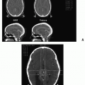
radiation insult in radiation injury to the central nervous system (CNS) is a demyelinating lesion with focal or diffuse areas of white matter necrosis (9).


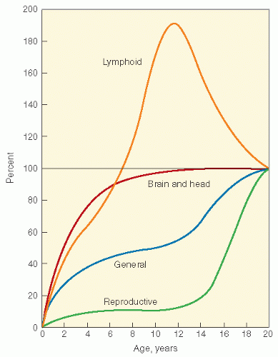
Figure 19.3 Growth curves of different tissues. (From Tanner JM. Growth at Adolescence. Oxford: Blackwell Scientific, 1962, with permission.)

The basic mechanisms underlying the pathologic changes are not precisely known for any particular syndrome of irradiation damage. The three most commonly proposed mechanisms may act alone or in combination. The vascular mechanism acknowledges that the endothelial cell is essential for patency of the microcirculation. This cell is radiosensitive, and damage is expressed as cell death or endothelial hyperplasia. Because endothelial cell turnover is slow, injury based on these cells occurs over a prolonged time interval. The evolution of hyalin degeneration and obliterating sclerosis of the arterioles produces areas of complete and incomplete necrosis. The clonogenic death of glial cells mechanism postulates a radiation-induced reproductive death of the slowly reproducing oligodendrocyte. The oligodendrocyte maintains myelin. These cells show a decrease in numbers within weeks after irradiation. Damage in individual nerve fibers can be demonstrated quantitatively by electron microscopy as early as 2 weeks after irradiation and before vascular damage. Effects on myelin synthesis and maintenance may be especially important in childhood because myelogenesis is most active in the first year of life. The ultimate result is demyelination. Radiation-induced endothelial cell death followed by vascular occlusion also promotes necrosis. The allergic mechanism of pathologic change in the brain after irradiation argues that the lesions of delayed radiation necrosis sometimes consist of disseminated plaques of demyelination with central necrosis and occasional petechial hemorrhage. In the patent blood vessels that remain, there may be perivascular cuffing with lymphocytes and plasma cells. The postulated autoimmune mechanism is that an antigen is produced by the reaction of ionizing irradiation with the oligodendromyelin complex. This antigen would stimulate the accumulation of inflammatory cells.

The classic findings of radiation-induced necrosis of the CNS are large areas of confluent, coagulative necrosis of the white matter and deep layers of the cortex; the vascular changes of fibrinoid necrosis or fibrin incontinence; atypia or absence of endothelial cells; vascular thickening; telangiectasis; and vascular proliferation.


Clinical Manifestations

Necrosis. The incidence of radiation necrosis after therapeutic dosages is uncertain. Quoted incidence data range from 0.1% to 5% after dosages of 50-60 Gy fractionated over 5-6 weeks (9,10). The variability in the data is the result of the uncertainty, in some studies, concerning the denominator; the long time delay until the occurrence of necrosis; the lack of histologic confirmation in many surviving patients; and the absence of postmortem data in patients who succumb. The clinical signs and symptoms of radiation necrosis are headache and mass effect. Surgical debulking is performed when possible and is often therapeutic (10). Corticosteroids may offer transient relief. Anecdotal reports of the benefits of anticoagulation with heparin or warfarin compounds exist (11,12).

Necrotizing Leukoencephalopathy and Mineralizing Microangiopathy. Leukoencephalopathy is a late complication of cranial irradiation and systemic methotrexate (MTX). The histologic appearance is of multifocal white matter destruction, especially in the centrum semiovale and periventricular regions, with loss of myelin and oligodendrocytes. Hypodense areas emerge in the white matter, and there is cerebral atrophy, an increase in the sulcal width, and enlargement of the ventricles. Mineralizing microangiopathy can occur and is visualized on computed tomography (CT) scanning as intracerebral calcification (11). The clinical features include lethargy, seizures, spasticity, paresis, and ataxia. The multifactorial origin and risk of leukoencephalopathy have been studied in acute lymphoblastic leukemia (ALL) survivors. Radiation and MTX appear to be contributing factors (Fig. 19.4) (13). Of interest is a report from the German Late Effects Working Group, which documented CT and magnetic resonance imaging (MRI) evidence of atrophy, leukoencephalopathy, calcification, or gray matter changes in approximately 50% of children treated for ALL (14). The frequency and severity of abnormalities were greater in children treated with cranial RT and MTX than in those treated with MTX alone. It is worth emphasizing that the situation is decidedly worse in children who have CNS involvement with ALL. In these cases, the presence of leukemia can alter the cerebrospinal clearance of MTX and increase the risk of injury.

Neuropsychologic and Intellectual Deficits. Cranial irradiation of children may have significant adverse effects on intelligence, learning, and social and emotional adjustment
(11,12,15, 16, 17). Neurocognitive late effects most commonly follow treatment of malignancies that necessitate CNSdirected therapies, such as cranial radiation, intraventricular or IT or high dose systemic (CNS penetrating) chemotherapy. Thus, children with CNS tumors, head and neck sarcomas, and ALL are most commonly affected. Studies of patients with brain tumors must account for the confounding variables of CNS injury by the tumor, seizures and their therapy, elevated intracranial pressure, and surgery. In patients with leukemia and brain tumors, the use of chemotherapy and the impact of the illness on body image and school attendance may complicate the issue.


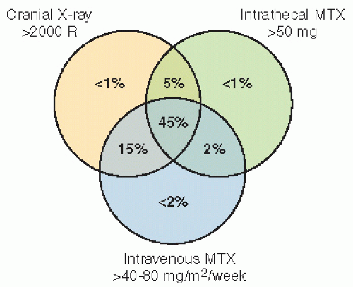
Figure 19.4 Approximate risks of leukocephalopathy (From Griffin TW. White matter necrosis, microangiopathy, and intellectual abilities in survivors of childhood leukemia. Association with central nervous system irradiation and methotrexate therapy. In: Gilbert HA, Kagan AR, eds. Radiation Damage to the Nervous System. New York: Raven Press; 1980:155-174, with permission).

Deficits occur in a variety of areas, which include general intelligence, age-appropriate developmental progress, academic achievement (especially in reading language and mathematics), visual and perceptual motor skills, nonverbal and verbal memory, receptive and expressive language, and attention (11,12,15, 16, 17). Younger age at the time of treatment is associated with a greater neurocognitive deficit (18).

The effects of cranial RT on subsequent intellectual function have been studied in detail, most comprehensively in survivors of CNS tumors and leukemia. However, the effects of radiation on the brain are difficult to define, especially when cranial radiation is often routinely a part of multimodality therapy that may also include surgery and systemic or IT chemotherapy. Moreover, tumor-related deficits caused by direct invasion of the brain, seizures, and hydrocephalus must be recognized.

With detailed assessment of IQ, achievement tests, learning ability, and school performance, it is clear that cranial RT can result in significant neurocognitive dysfunction. With changes in the dosage, fields, and volumes of cranial and craniospinal RT, changes in the incidence and spectrum of these adverse effects of therapy are expected. More recent studies using lower dosages and more targeted volumes have demonstrated better results (19, 20, 21). Ris et al. (19) evaluated the intellectual outcome of 43 children treated on Children’s Cancer Group study 9892 for posterior fossa medulloblastoma or primitive neuroectodermal tumor (PNET). The study used a reduced craniospinal RT regimen of 23.4 Gy to the neuraxis and 32.4-Gy boost to the posterior fossa with adjuvant chemotherapy. The mean age at the time of treatment was 6 years, with a range of 3-15 years. The estimated rate of change of FSIQ was 4.3 points/year, for VIQ 4.2 points/year, and for nonverbal IQ (NVIQ) 4.0 points/year. In examining host-related factors, a more significant decline was seen for girls (-8 points/year) than boys (-3 points/year) for VIQ and for children less than 7 years old (-5.2 points/year) as opposed to children 7 years and older (-0.8 points/year) for NVIQ. This latter observation was noteworthy in that the older children’s baseline was lower, with little decline over time, as compared with the younger children, who had a much higher baseline followed by a steep decline. The effect of baseline intelligence was examined for the whole group, and the same phenomenon was seen. Children who had a baseline IQ of 100 or higher had a much steeper decline in FSIQ, VIQ, and NVIQ than children who had a baseline under 100. The effect on FSIQ is shown in Figure 19.5.


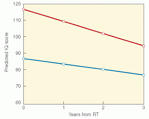
Figure 19.5 Full-scale IQ change by baseline intelligence. Blue, baseline IQ < 100; Red, baseline IQ > 100. (From Ris MD, Packer R, Goldwein J, et al. Intellectual outcome after reduceddose radiation therapy plus adjuvant chemotherapy for medulloblastoma: a Children’s Cancer Group study. J Clin Oncol. 2001;19:3470-3476, with permission.)

Another recent study supports the hypothesis that patients with medulloblastoma demonstrate a decline in IQ values because of an inability to acquire new skills and information at a rate comparable to that of their healthy same-age peers rather than a loss of previously acquired information and skills (15). In a Danish study of 133 children treated for brain tumors, younger age at diagnosis, tumor site in the cerebral hemisphere, hydrocephalus treatment with shunt, and radiation therapy predicted lower cognitive functions (22). Finally, a reduction in the volume of normal-appearing white matter has been observed in some survivors of childhood brain tumors, and this was associated with decreased attentional abilities, which led to an IQ deficit and impaired academic achievement (16).

For ALL, older studies again show significant neurocognitive impairment (23). Even when combined with IT chemotherapy, reduction in the cranial radiation dosage has resulted in less neurocognitive impairment (18,24,25). Studies on CNS prophylaxis for ALL comparing craniospinal
RT with cranial RT combined with IT MTX showed that children who were less than 5 years old at the time of treatment and had received RT and IT chemotherapy had lower IQ scores than those who received craniospinal RT alone. Meadows et al. (23) found a significant IQ deficit in children treated with 24-Gy cranial RT combined with IT MTX, as compared with childhood cancer survivors who received no CNS-directed therapy, with the effect greatest among those less than 5 years old. A similar effect on cognition with the addition of IT MTX has been found in children treated for medulloblastoma (26).

Controversy regarding the role of RT in impairing neurocognition will continue, largely because of differences in the radiation dosages and drugs used in various protocols and different endpoints. Prophylactic cranial irradiation of 24 Gy at 2 Gy per fraction in the treatment of ALL can lower IQ and achievement test scores. IQ loss may be on the order of 8-10 points (i.e., median of normals of 100 down to 90-92). Some studies suggest that the undesirable consequences of 18 Gy are less than 24 Gy, and that 12 Gy (commonly used in current protocols) does not cause identifiable deficits, but the data on this point are not definitive (23).

The deleterious effects of systemic MTX, especially at dosages greater than 1 g/m2, may be no different from those of 18-Gy cranial RT (27,28). At lower MTX dosages, there does not appear to be a consistent pattern of neurocognitive deficits (29). A recent long-term study of infants who received high-dose systemic MTX combined with IT cytarabine and MTX for CNS leukemia prophylaxis and were tested 3-9 years after treatment showed that cognitive function was in the average range (30).

In a small study from Italy, cognitive outcome was evaluated in 21 children who received both cranial RT and IT MTX. Mean total IQ was within normal limits, but there was a large discrepancy between the VIQ and PIQ, with the former being significantly higher than the latter. In addition, intracerebral calcifications on MRI were associated with the number of intrathecal MTX dosages and with low scores on IQ, attention, and visual integration tests. Age at treatment and RT dosage were not associated with neuropsychiatric outcomes (31).

Chemotherapy without RT for ALL may result in cognitive dysfunction, but study results vary significantly. Some studies have demonstrated impairment in tasks of higherorder cognitive functioning, mathematics (27), and mild visual and verbal short-term memory deficits (32) in leukemia survivors treated with intrathecal chemotherapy. Yet, others have demonstrated no significant problems (24,33,34). The substitution of dexamethasone for prednisone in the treatment of ALL has been implicated in increasing cognitive dysfunction (25), but a recent study examining differences between children treated for ALL on a study, where there was a randomization between prednisone and dexamethasone, found no significant differences in neurocogntive performance (Kadan-Lottick) (34).

Somnolence Syndrome. Irradiation of the brain of children can result in the somnolence syndrome. The syndrome has its onset 4-8 weeks after irradiation and is characterized by drowsiness, nausea, irritability, anorexia, apathy, and dizziness. The majority of children reported to have developed this syndrome were irradiated for ALL, although it can occur after treatment of a brain tumor. The somnolence syndrome resolves spontaneously, but corticosteroids hasten recovery.

Arterial vasculopathy is an uncommon occurrence, almost always described after irradiation to the parasellar region, primarily in children. Single- or multiple-vessel narrowing or obliteration results in typical stroke deficits (35).

Myelopathy. The spectrum of radiation injuries to the spinal cord includes transient and irreversible syndromes. A rare, rapidly evolving permanent paralysis is presumed to result from an acute infarction of the cord. A more common form of radiation injury is manifested by the Lhermitte sign or Lhermitte symptom, which consists of an electric shock-like sensation that radiates down the spine and often into the limbs (36). The location of the sensation can change with time. The symptoms may be precipitated by flexion of the neck, walking on a hard surface, sitting on a hard surface, or other forms of physical exertion. The syndrome generally occurs a few months after spinal irradiation. The mechanism is thought to be transient demyelinization, although detailed human pathologic studies are lacking. There are no known CT or MRI correlations. It is also reported in association with cisplatin administration, where the results may be long-lasting (37).

Chronic progressive radiation myelitis (CPRM) is rare. The initial symptoms, generally subtle, are usually paresthesias and sensory changes (including diminished temperature sensation or proprioception), which start 9-15 months after therapy and progress over the subsequent year (38). Much longer intervals to initial symptoms are seen occasionally. The neurologic lesion must be within the irradiated volume. Recurrent or metastatic tumor must be ruled out. Cerebrospinal fluid (CSF) protein may be elevated, and myelography can demonstrate cord swelling or atrophy. MRI and CT provide additional supportive information.

The incidence of CPRM and the radiation dosage causing this event are poorly defined because of the diagnostic difficulties and the variety of radiation techniques (with uncertain dosimetries). A review by Wara et al. (39) suggests that 42 Gy in 25 fractions carries a 1% risk, 45 Gy a 5% risk, and 61 Gy a 50% risk. Data from Marcus and William (40) suggest that the cervical cord may be more tolerant to radiation than previously presumed when 1.8- to 1.9-Gy fractional doses are used; in 324 patients treated to dosages of 55 Gy or less, no cases of myelitis were seen. A higher risk of myelopathy is associated with higher individual fraction sizes, shorter overall treatment time, higher total dosages, and long lengths of the cord treated (especially more than 10 cm). Children may be more susceptible to CPRM, developing it after lower radiation dosages and with shorter latency periods (38).

Table 19.3 reviews several CNS late effects with respect to causative treatments, signs and symptoms, screening and diagnostic tests, and management and intervention (41).


Psychosocial

Psychological Effects. Due to the lack of systematic screening for patients with and without evident psychsocial difficulties, the prevalence of psychological distress among survivors is poorly characterized. It is clear, however, that a meaningful minority of childhood cancer survivors and their parents demonstrate symptoms of uncertainty, distress and posttraumatic stress, with some meeting full criteria for posttraumatic stress disorder (PTSD). Because avoidance of

places and people associated with the cancer is part of PTSD, the syndrome may interfere with appropriate access to health care. Risk factors include perception of threat to life and severity of treatment, poor family functioning and decreased social support (42, 43, 44, 45, 46, 47).


Table 19.3 Evaluation of Patients at Risk for Late Effects: Central Nervous System


Causative Treatment


Signs and Symptoms


Screening and Diagnostic Tests


Management and Intervention


Late Effects


Chemotherapy


Radiation


Surgery


Neurocognitive deficit


High-dose IV MTX, IT MTX


>18 Gy


Resection of central nervous system tumor


Difficulty with reading, language, verbal and nonverbal memory, arithmetic, receptive and expressive language, decreased speed of mental processing, attention deficit, decreased IQ, behavior problems, poor school attendance, poor hand-eye coordination


Neurocognitive testing: Psychoeducational Neuropsychologic


Psychoeducation assistance


Leukoencephalopathy


IT or IV MTX


IT cytosine arabinoside


>18 Gy (with MTX)


Seizures, neurologic impairment; compare with premorbid status


CT and MRI for baseline and symptoms


Symptom management: muscle relaxants, anticonvulsants, physical therapy, occupational therapy


Focal necrosis


IT or high-dose IV MTX


Carmustine, cisplatin


>50 Gy (especially with >2 Gy daily fraction)


Tumor resection


Headaches; nausea; seizures; papilledema hemiparesis or other focal findings; speech, learning, and memory deficits


CT and MRI for baseline, PRN


Positron emission tomography or single-photon emission computed tomography


Steroid therapy


Debulking of necrotic tissue


Large-vessel stroke


>35 Gy


Headache, seizures, hemiparesis, aphasia, focal neurologic findings


CT and MRI Arteriogram


Determined by specific neurologic impairment


Vision loss


Intra-arterial carmustine, cisplatin


>50 Gy (optic nerve chiasm, occipital lobe)


Tumor resection


Progressive visual loss


Ophthalmic evaluation


Visual-evoked response


Visual aids


Ototoxicity


Cisplatin, carboplatin


>35 Gy (middle and inner ear)


Surgery, CSF shunting


Abnormal speech development Hearing


Audiogram for baseline, PRN


Speech therapy Hearing aid


Myelitis


45-50 Gy


Spinal cord surgery


Paresis, spasticity, altered sensation, loss of sphincter control


MRI


Steroids


Physical therapy


Occupational therapy


IT, intrathecal; MTX, methotrexate; IV, intravenous; CT, computed tomography; MRI, magnetic resonance imaging; PRN, as needed; CSF, cerebrospinal fluid.


From Constine LS, Hobbie W, Schwartz C. Facilitated assessment of chronic treatment by symptom and organ systems. In: Schwartz C, Hobbie W, Constine L, et al., eds. Survivors of Child and Adolescent Cancer: A Multi-Disciplinary Approach. Berlin: Springer-Verlag; 2005:17-34, with permission.


A combined analysis of symptoms of somatic distress and depression has been completed from the Childhood Cancer Survivor Study (CCSS) cohort. This included 1834 survivors of HL who were compared with sibling controls. Eighty-seven had scores on the brief symptom index that were symptomatic for depression, and 237 for somatic distress. Risk factors included female sex, low socioeconomic status, lack of a complete high school education, lack of employment, intensive chemotherapy, and elapsed time since therapy (48). In a subsequent analysis of the entire CCSS cohort, Mulrooney et al. (49) evaluated risk factors for fatigue or sleep impairment. In univariate analysis, compared with other diagnostic groups, a diagnosis of HL or sarcoma was associated with higher risks of fatigue, depression, and sleep impairment. In multivariate analysis of the fatigue risk factors, hypothyroidism and depression were independent risk factors, and radiation therapy had a marginal association.


Neuroendocrine

Hypothalamic-pituitary (HP) irradiation may produce a spectrum of neuroendocrine abnormalities.


Growth Hormone

The effects of cranial irradiation on growth hormone (GH) production and release are most common. GH is secreted episodically by the anterior pituitary gland. Growth hormone-releasing hormone (GHRH), produced in the hypothalamus, stimulates GH production, whereas the hypothalamic neuropeptide somatostatin excretes an inhibitory effect. In the liver and other tissues, circulating GH stimulates the production of insulin-like growth factor 1 (IGF-1, also called somatomedin-C), which promotes cell proliferation and protein synthesis. The anatomic site of the radiation injury that produces GH deficiency is the hypothalamus.


Clinical Manifestations

The potential for neuroendocrine damage is likely to decrease with the use of more focused RT and a decrease in dosage in some settings such as medulloblastoma. Approximately 60-80% of irradiated pediatric brain tumor patients who have received dosages greater than 30 Gy have impaired serum GH response to provocative stimulation. This usually occurs within 5 years of treatment. The dose-response relationship has a threshold of 18-20 Gy, and the higher the radiation dosage, the earlier the GH deficiency will occur after treatment. A study of conformal RT in children with CNS tumors indicates that GH insufficiency usually can be demonstrated within 12 months of RT depending on hypothalamic dosevolume effects (50). Children treated with CNS irradiation for leukemia are also at greater risk of GH deficiency. Sklar et al. (51) evaluated 127 patients with ALL treated with 24 Gy, 18 Gy, or no cranial irradiation. The change in height standard deviation score (SDS) was significant for all three groups, with a dose-response relationship noted: 0.49-0.14 for no RT group, 0.65-0.15 for the 18-Gy RT group, and 1.38-0.16 for the 24-Gy group. In the CCSS, Chow demonstrated that risk for adult short stature is highest among patients treated with cranial or craniospinal RT, but is also elevated among those treated with chemotherapy alone (52). Figure 19.6 shows the height SDS for ALL survivors, treated pre- or postpubertally with chemotherapy alone, cranial or craniospinal RT as compared with siblings.


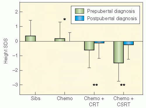
Figure 19.6 Height standard deviation scores (SDS) for ALL survivors, treated pre- or postpubertally with chemotherapy alone, cranial or craniospinal RT. (From Chow EJ, Friedman DL, Yasui Y, et al. Decreased adult height in survivors of childhood acute lymphoblastic leukemia: a report from the Childhood Cancer Survivor Study. J Pediatr. 2007;150:370-375, with permission)

Children who receive bone marrow transplantation (BMT) with total body irradiation (TBI) have a significant risk of GH deficiency. Risk is higher with single-dose as opposed to fractionated radiation, pretransplant cranial irradiation, female sex, and posttreatment complications such as graft-versus-host disease (GVHD) (53,54). Regimens with busulfan and cyclophosphamide also increase risk (54). Hyperfractionation of the TBI dosage markedly reduces risk in the absence of pretransplant cranial radiation (55). In a review, Socie et al. (56) discuss this risk at length. The mean loss of height is estimated to be approximately 1 SDS (6 cm) compared with the mean height at the time of stem cell transplantation and mean genetic height. In a report from the European Group for Blood and Marrow Transplantation, among 181 patients with aplastic anemia, leukemias, and lymphomas who underwent stem cell transplantation before puberty, an overall decrease in final height SDS value was found compared with the height at transplant and with the genetic height. The type of transplantation, GVHD, GH, or steroid treatment did not influence final height. TBI (single greater than fractionated dosage), male sex, and young age at transplant were found to be major factors for long-term height loss. The majority of patients (140 of 181) reached adult height within the normal range of the general population. Data from this study demonstrating the changes in SDS by type of RT are shown in Figure 19.7 (57).

GH deficiency should be treated with replacement therapy. There has been some controversy surrounding this therapy, with a concern over increased risk of recurrence and second malignant neoplasms (SMNs). However, most studies are limited by selection bias and small sample size. Sklar et al. (58) studied 361 GH-treated cancer survivors enrolled in the
CCSS and compared risk of recurrence, risk of SMN, and risk of death between survivors who did and did not receive treatment with GH. The relative risk of disease recurrence was 0.83 (95% CI 0.37-1.86) for GH-treated survivors. GHtreated subjects were diagnosed with 15 SMNs, all solid tumors, for an overall relative risk of 3.21 (95% CI 1.88-5.46), mainly because of a small excess number of SMNs observed in survivors of acute leukemia. Two other studies have shown no increased risk of relapse or SMN (59,60). The data surrounding SMNs must be interpreted with caution given the small number of events.


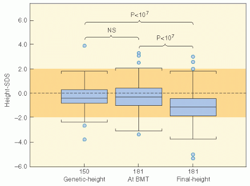
Figure 19.7 Correlation between the genetic height, height standard deviation score (SDS) at bone marrow transplantation, and final height SDS. Numerals indicate the number of cases studied in each group. The dotted area indicates the height SDS distribution for the normal general population. The lower line in the box plot indicates the 25th percentile, the upper line indicates the 75th percentile, and the horizontal lines above and below the boxes represent the 3rd and the 97th percentile, respectively. Statistical analyses: paired Student’s t test. (From Cohen A, Rovelli A, Bakker B, et al. Final height of patients who underwent bone marrow transplantation for hematological disorders during childhood: a study by the Working Party for Late Effects-EBMT. Blood. 1999;93:4109-4115, with permission.)


Non-Growth Hormone Tropins

Pubertal growth can also be adversely affected by cranial radiation. Dosages greater than 50 Gy may result in gonadotropin deficiency, and dosages of 18-47 Gy can result in precocious puberty. Precocious puberty has occurred mostly in girls who received dosages of at least 24 Gy cranial radiation. However, earlier puberty and earlier peak height velocity are seen in girls treated with 18 Gy cranial radiation (61).

Shalet et al. (62) showed that the age of pubertal onset is positively correlated with the age at the time of cranial irradiation. The impact of early puberty in a child with radiation-associated GH deficiency is significant, and the timing of GH is especially important for GH-deficient girls also at risk of precocious puberty. With higher dosages of cranial irradiation (more than 35 Gy), deficiencies in the gonadotropins can be seen, with a cumulative incidence of 10-20% at 5-10 years (63).

Constine et al. (64) documented non-GH abnormalities in 20 children treated with irradiation for brain tumors not involving the HP region, including low free T4 levels caused by hypothalamic or pituitary injury and low luteinizing hormone (LH) and estradiol with oligomenorrhea. The frequency of central hypothyroidism after cranial irradiation relates to the dosage to the HP axis, with a greater likelihood after dosages greater than 40 Gy. Although some reports suggest that the incidence is as low as 6%, the radiation dosages to the HP axis in those reports were not specifically determined (65,66). In Constine et al.’s series (67), 65% of patients treated in the higher dosage range had evidence of subclinical or clinical hypothyroidism. In a report by Paulino (68) on children treated for medulloblastoma, and thus with somewhat lower HP dosages, 19% of children developed central hypothyroidism.

Adrenocorticotropin deficiencies and hyperprolactinemia are rare in children because dosages greater than 50 Gy are needed for their development (64,69). Samaan et al. (70) described 110 patients treated for nasopharyngeal or paranasal sinus carcinoma, in whom more than 60 Gy was typically administered. They found deficiencies in 83%. Twenty-seven percent had thyroid abnormalities, attributed to hypothalamic injury in one third of the patients and pituitary injury in the remainder. Cortisol deficiency attributable to hypothalamic injury also occurred in 27% of patients. Abnormalities of prolactin and LH were noted in 39% and 30%, respectively. A summary of neuroendocrine complications of therapy, the relationship to dosage, diagnostic studies, and interventions are provided in Table 19.4 (41).


Thyroid

Radiation-induced thyroid neoplasms are considered in Chapter 20. In this section, we will review the effects of therapeutic irradiation on the endocrine function of the thyroid. The thyroid may be directly irradiated in the treatment of childhood head and neck cancers such as rhabdomyosarcoma, nasopharyngeal lymphoepithelioma, and squamous cell carcinoma, a variety of other aerodigestive tract tumors, and HL. A photon spinal field used during craniospinal irradiation for brain and spinal tumors and for leukemia will deliver radiation to the thyroid.


Pathophysiology

The thyroid gland consists of follicles filled with colloid and lined by follicular cells, which trap iodide. The glycoprotein thyroglobulin is a major component of the colloid and participates in the formation and storage of thyroid hormones. The primary, hormonally active iodothyronines,


triiodothyronine (T3) and thyroxine (T4), are largely bound to plasma proteins when released by the gland. Thyroxinebinding globulin is the major transport protein, and only a small percentage of unbound T3 and T4 is available for activity. The pituitary hormone thyroid-stimulating hormone (TSH) regulates synthesis and release of T3 and T4. TSH secretion is stimulated by the hypothalamic hormone thyrotropin-releasing hormone (TRH) and inhibited by the circulating free thyroid hormones. The pathophysiology of radiation-induced thyroid dysfunction is not precisely defined. Direct radiation damage to the thyroid follicular cells, the thyroid vasculature, or the supporting stroma may occur. Less likely mechanisms that could contribute include the induction by RT of an immunologic reaction or damage from the iodine load administered for lymphangiography. Histopathologic changes in an irradiated thyroid gland include progressive obliteration of the fine vasculature, degeneration of follicular cells and follicles, atrophy of the stroma, and, less commonly, lymphocytic infiltration. Because radiation damage depends on the degree of mitotic activity and the thyroid of a developing child grows in parallel with body growth, this gland might be expected to show an age-related degree of injury and repair.


Table 19.4 Causative Treatment


Causative Treatment


Signs and Symptoms


Screening and Diagnostic Tests


Management and Intervention


Late Effects


Chemotherapy


Radiation


Surgery


GH deficiency


>18 Gy to HP axis


Tumor in region of HP axis


Falling off of growth curve


Inadequate growth velocity


Inadequate pubertal growth spurt


Annual stadiometer height (q6 months at age 9-12 years)


Growth curve


Bone age at 9 years, then yearly until puberty is reached


Insulin stimulation test and pulsatile GH analysis


GH therapy


Delay puberty with GnRH agonist


Adrenocorticotropic hormone deficiency


>40 Gy to HP axis


Tumor in region of HP axis


Muscular weakness, anorexia, nausea, weight loss, dehydration hypotension, abdominal pain, increased pigmentation (skin, buccal mucosa


Cortisol (a.m.) for baseline, PRN


Insulin—hypoglycemia; metapyrone stimulation tests


Hydrocortisone


Thyrotropinreleasing hormone deficiency


>40 Gy HP axis


Tumor in region of HP axis


Hoarseness, fatigue, weight gain, dry skin, cold intolerance, dry brittle hair, alopecia, constipation, lethargy, poor linear growth, menstrual irregularities, pubertal delay, bradycardia, hypotension


Free T4, T3, TSH baseline, ql year


Hormone replacement with thyroxine


Anticipatory guidance regarding symptoms of hypothyroidism


Precocious puberty (especially girls)


>20 Gy to HP axis


Tumor in region of HP axis


Early growth spurt


False catch-up


Premature sexual maturation:


Female: Breast development and pubic hair before 8 years and menses before 9 years


Male: testicular and penile growth and pubic hair before 9.5 years


Height, growth curve q yr


Bone age q2 years until mature


LH, FSH, estradiol, or testosterone


Pelvic ultrasound, GnRH stimulation testing


GnRH agonist


Male gonadotropin deficiency


>40 Gy to hypothalamic region


Tumor in region of hypothalamus


Delayed, arrested, or absent pubertal development: lack of or diminished pubic and axillary hair, penile and testicular enlargement, voice change, body odor, acne


Testicular atrophy (softer and smaller)


Failure to impregnate


LH, testosterone ql-2 years GnRH testing


Testosterone replacement


Female gonadotropin deficiency


>40 Gy to hypothalamic region


Tumor in region of hypothalamus


Delayed, arrested, or absent pubertal development, including breasts, female escutcheon, female habitus, vaginal estrogen effect, body odor


Tanner stage


Anticipatory guidance regarding symptoms of estrogen deficiency


Acne


Changes in duration, frequency, and character of menstruation (less cramping)


Estrogen deficiency: hot flashes, vaginal dryness, dyspareunia, low libido Infertility


If on birth control pills, then adjust dose


LH, FSH, estradiol ql-2 years


GnRH stimulation tests


Hormone replacement


Early intervention may prevent osteoporosis, atherosclerosis


Hyperprolactinemia


>40 Gy HP axis


Tumor in region of hypothalamus


Female: menstrual irregularities, loss of libido, galactorrhea, hot flashes, osteopenia


Male: loss of libido, impotence, infertility


Prolactin level baseline, then PRN


Dopamine agonist (bromocriptine)


Metabolic syndrome


Steroids


?≥18 Gy (dosage not well established)


Obesity, hypertension, hyperlipidemia, hyperglycemia, insulin resistance with hyperinsulinemia


Fasting lipids, glucose, insulin levels, body mass index evaluation


Refer to endocrinology


GH, growth hormone; HP, hypothalamic-pituitary; GnRH, gonadotropin-releasing hormone; PRN, as needed; LH, luteinizing hormone; FSH, follicle-stimulating hormone; TSH, thyroid stimulating hormone.


From Constine LS, Hobbie W, Schwartz C. Facilitated assessment of chronic treatment by symptom and organ systems. In: Schwartz C, Hobbie W, Constine L, et al., eds. Survivors of Child and Adolescent Cancer: A Multi-Disciplinary Approach. Berlin: Springer-Verlag; 2005:17-34, with permission.


Hypothryoidism

Of children treated with radiation therapy, most develop hypothyroidism within the first 2-5 years posttreatment, but new cases can occur later. Reports of thyroid dysfunction differ depending on the dose of radiation, the length of follow-up, and the biochemical criteria utilized to make the diagnosis. The most frequently reported abnormalities include elevated TSH, depressed thyroxine (T4), or both (71, 72, 73, 74). Compensated hypothyroidism includes an elevated TSH with a normal T4 and is asymptomatic. Uncompensated hypothyroidism includes both an elevated TSH and a depressed T4.

The incidence of hypothyroidism should decrease with lower cumulative doses of radiation therapy employed in newer protocols. In a study of Hodgkin lymphoma (HL) patients treated between 1962 and 1979, hypothyroidism occurred in 4 of 24 patients who received mantle doses less than 26 Gy but in 74 of 95 patients who received greater than 26 Gy. The peak incidence occurred at 3-5 years post treatment, with a median of 4.6 years (67). A cohort of childhood HL survivors treated between 1970 and 1986 were evaluated for thyroid disease by use of a self-report questionnaire in the CCSS. Among 1791 survivors, 34% reported that they had been diagnosed with at least one thyroid abnormality. For hypothyroidism, there was a clear dose response, with a 20-year risk of 20% for those who received less than 35 Gy, 30% for those who received 35-44.9 Gy, and 50% for those who received greater than 45 Gy to the thyroid gland. The RR for hypothyroidism was 17.1. Elapsed time since diagnosis was a risk factor, where the risk increased in the first 3-5 years after diagnosis (71). The cumulative incidence of hypothyroidism among survivors 15 years following leukemia diagnosis has also been evaluated in the CCSS and was 1.6%. In multivariate analysis, survivors who received ≥20 Gy cranial RT plus any spinal RT had the highest risk of subsequent hypothyroidism (HR 8.3) compared with those treated with chemotherapy alone. In radiation dosimetry analysis, pituitary doses ≥20 Gy combined with thyroid doses ≥10 Gy were associated with hypothyroidism (75). Figure 19.8 shows the cumulative incidence of hypothyroidism respectively after treatment for HL.


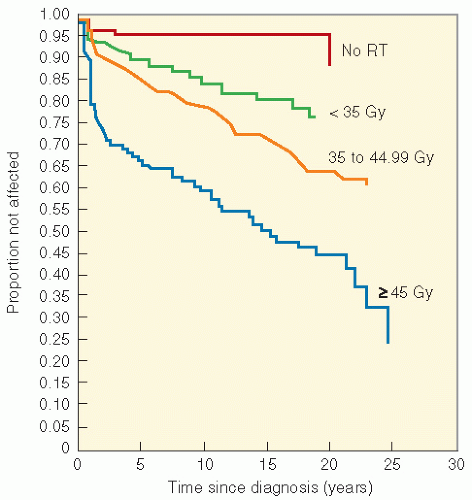
Figure 19.8 Probability of developing an underactive thyroid after diagnosis of the Hodgkin disease. (From Diller L, Chow EJ, Gurney JG, et al. Chronic disease in the Childhood Cancer Survivor Study Cohort: a review of published finding. J Clin Oncol. 2009;27:2339-2355.)

Survivors of pediatric hematopoietic stem cell transplantation (HSCT) are at increased risk of thyroid dysfunction, with the risk being much lower (15-16%) after fractionated TBI, as opposed to single-dose TBI (46-48%). While mildly elevated TSH is common, it is usually accompanied by normal thyroxine concentration (76). Non-TBI-containing regimens historically were not associated with an increased risk. However, in a recent report from the Fred Hutchinson Cancer Research Center, the increased risk for thyroid dysfunction was not different between children receiving a TBI or busulfan-based regimen compared with cylophosphamide conditioning alone.


Hyperthyroidism

Thyrotoxicosis may occur after mantle or cervical irradiation for HL. In Hancock et al.’s report (77), approximately 2% of patients (n = 34) developed Graves disease. Almost all had a diffuse goiter, high free T4, low TSH, and elevated thyroid uptake of radioiodine. One half of these patients developed infiltrative ophthalmopathy, as did an additional four patients who did not have overt hyperthyroidism. The relative risk for Graves disease was 7.2-20.4. Six patients developed silent thyroiditis characterized by transient mild symptoms of thyrotoxicosis, elevated serum-free T4 and low TSH, no thyroid enlargement or tenderness, and low thyroid uptake of radioiodine (78). All of these patients subsequently developed hypothyroidism.

In the CCSS study of HL survivors by Sklar et al. (71), the relative risk for hyperthyroidism compared with sibling control subjects was 8.0, with cases becoming manifest
3-5 years after diagnosis. In the study of ALL survivors from the CCSS, the 15-year cumulative incidence was 0.6%. Craniospinal RT was associated with an increased risk of subsequent hyperthyroidism (HR 6.1) compared with chemotherapy alone. In radiation dosimetry analysis, pituitary doses ≥20 Gy combined with thyroid doses ≥15 Gy were associated with hyperthyroidism (75).


Detection and Screening

Laboratory screening evaluations for asymptomatic patients should include serum concentrations of TSH and free thyroxine. Because the latent interval to the development of abnormality can be prolonged, systematic clinical and laboratory evaluation should be performed yearly.


Management

Patients with uncompensated hypothyroidism (low serum concentration of thyroxine) clearly need thyroid replacement therapy. Patients with elevated serum concentrations of TSH but normal thyroxine are treated with thyroid replacement therapy in most institutions. The rationale for this approach is that subclinical hypothyroidism may evolve into overt hypothyroidism, and prolonged TSH stimulation of an irradiated thyroid gland may increase the risk of carcinoma (67,77).

Table 19.5 reviews several thyroid late effects with respect to causative treatments, signs and symptoms, screening and diagnostic tests, and management and intervention (80).


Bone and Body Composition


Pathophysiology

It has long been established that bone growth can be impaired by radiation. The pathophysiology of radiation injury to growing bone probably is attributable to damage to the chondroblasts. Single doses of 2-20 Gy inhibit proliferation of cartilage cells, thereby decreasing cellularity and causing disarray of the cellular columns within the growth plate. The resulting retarded bone growth is attributed to this loss of proliferating cells in the growth plate, the decreased ability of surviving cells to synthesize matrix, or the production of an abnormal matrix that fails to calcify.

The effects of radiation on growing bone have been summarized by Rubin et al (6). Epiphyseal irradiation, to a sufficient dosage, causes an arrest of chondrogenesis; metaphyseal irradiation results in a failure of absorptive processes in calcified bone and cartilage; and diaphyseal irradiation produces an alteration in periosteal activity, which causes abnormal bone modeling. Therefore, the location of an RT field on a long bone can significantly influence the nature and severity of the subsequent deformity.


Clinical Manifestations

Bone Growth Retardation. Chondroblasts and chondrocytes are affected by radiation therapy in growing children, and this can result in soft tissue hypoplasia and diminution of bone growth. These effects are associated with the total and fractional radiation dosage and the inclusion of the epiphyses in the radiation field.

Clinically, radiation’s effects on growing bone may be most simply characterized as shortening of long bones (i.e., femur, tibia, humerus) or hypoplasia of flat bones (i.e., ilium). The crucial factors influencing the ultimate height of the patient may be inferred from the data previously presented: the radiation total dosage and dosage per fraction; the energy, dosage distribution, and absorptive properties of the beams; the bones that are irradiated and the epiphyseal plates that are encompassed; the age at the time of irradiation (implying that the amount of growth already obtained is important in judging ultimate outcome); the influence of other toxins on growth, such as exogenous steroids and cytotoxic chemotherapy; and the patient’s genetic constitution.

A large body of knowledge has been developed concerning loss of height after childhood irradiation. Much of the data published in the 1970s and 1980s reflect treatment with higher dosages of RT than those currently used. However, several recent reports indicate that stature loss must still be considered in the late effects of RT. For children treated with craniospinal RT for ALL or CNS tumors, the cranial dosage can result in decreased growth by causing growth hormone deficiency, and the spinal dosage can result in stature loss through its effect on the vertebral bodies (52).

Determining final adult height is not clear. Silber et al. (79) have attempted to address this problem with a mathematical model to predict adult stature after treatment of cancer in childhood. However, the model was based on data from only 49 patients. Donaldson (80) was unable to confirm the model’s efficacy in two of three cases.

Patients who receive radiation therapy for the Wilms tumor are an ideal population for study of the effect of RT and age at the time of treatment on stature. As a result of earlier studies showing a higher risk of spinal curvature with asymmetric radiation to the vertebral bodies, standard flank radiation now includes the affected side and extends across the spinal column but excludes the contralateral kidney. Radiation dosages have decreased over successive National Wilms Tumor Study Group (NWTSG) protocols. Hogeboom et al. (81) evaluated stature loss in 2778 children treated on NWTS 1-4. Repeated height measurements were collected during long-term follow-up. The effects of radiation dosage, age at treatment, and chemotherapy on stature were analyzed using statistical models that accounted for the normal variation in height with sex and advancing age. Predictions from the model were validated by descriptive analysis of heights measured at the age of 17-18 years for 205 patients. For those under 12 months of age at diagnosis who received more than 10 Gy, the estimated adult height deficit was 7.7 cm when contrasted with the nonradiation group. For those who received 10 Gy the estimated trunk shortening was 2.8 cm or less. Among those whose height measurements in the teenage years were available, patients who received more than 15 Gy of RT were 4-7 cm shorter on average than their nonirradiated counterparts, with a dose-response relationship evident. Chemotherapy did not confer additional risk.

Scoliosis and Kyphosis. Scoliosis and kyphosis are common consequences of spinal or flank irradiation. Asymmetric spinal growth has been seen most often after irradiation for the Wilms tumor and neuroblastoma, where there has been flank surgery (e.g., nephrectomy) and radiation (82). The types of deformities observed included a lateral flexion curve, concave to the side of the primary tumor, and a rotary scoliosis. However, with current dosages and fields of RT used to treat the Wilms tumor, scoliosis and kyphosis are less common. In a

series by Paulino et al. (83), in a group of 42 children treated for the Wilms tumor from 1968 to 1994, 7 developed muscular hypoplasia, 5 were found to have limb length inequality, 3 had kyphosis, and another 3 had iliac wing hypoplasia. Scoliosis was seen in 18, with only 1 patient needing orthopedic intervention. Median time to development of scoliosis was 102 months (range 16-146). A clear dose-response relationship was seen, with children treated with lower dosages (less than 24 Gy) having a significantly lower incidence of scoliosis than those who received more than 24 Gy. There was also a suggestion that the incidence was lower in patients who received 10-12 Gy, the dosages currently used for the Wilms tumor, although the sample size was small.


Table 19.5 Evaluation of Patients at Risk for Late Effects: Thyroid


Causative Treatment


Signs and Symptoms


Screening and Diagnostic Tests


Management and Intervention


Late Effects


Chemotherapy


Radiation


Surgery


Overt hypothyroidism (elevated TSH, decreased T4)


>20 Gy to the neck, cervical spine


>7.5 Gy total body irradiation


Partial or complete thyroidectomy


Hoarseness, fatigue, weight gain, dry skin, cold intolerance, dry brittle hair, alopecia, constipation, lethargy, poor linear growth, menstrual irregularities, pubertal delay, bradycardia, hypotension


Free T4, TSH annually up to 10 year postradiation or if symptomatic


Plot on growth chart


Refer to endocrinologist


Thyroxine replacement


Anticipatory guidance regarding symptoms of hyperthyroidism and hypothyroidism


Compensated hypothyroidism (elevated TSH, normal T4)


Same as for overt hypothyroidism


Same as for overt hypothyroidism


Asymptomatic


Same as for overt hypothyroidism


Refer to endocrinologist


Thyroxine tosuppress gland activity


Thyroid nodules


Any dosage


Same as for overt hypothyroidism


Same as for overt hypothyroidism


Physical exam


Ultrasound for technetium-99m scan for baseline and then PRN


Refer to endocrinologist


Thyroid scan


Biopsy or resection


Hyperthyroidism (low TSH, elevated T4)


Same as for overt hypo thyroidism


Nervousness, for tremors, heat intolerance, weight loss, insomnia, increased appetite, diarrhea, moist skin, tachycardia, exophthalmus, goiter


Same as thyroid nodules


T3, antithyroglobulin, antimicrosomal antibody baseline, then PRN


Refer to endocrinologist


PTU, propranolol


131I Thyroidectomy


TSH, thyroid-stimulating hormone; PRN, as needed; PTU, propothiouracil.


From Constine LS, Hobbie W, Schwartz C. Facilitated assessment of chronic treatment by symptom and organ systems. In: Schwartz C, Hobbie W, Constine L, et al., eds. Survivors of Child and Adolescent Cancer: A Multi-Disciplinary Approach. Berlin: Springer-Verlag; 2005:17-34, with permission.


Asymmetric irradiation of the vertebrae seemed to promote the development of rotary scoliosis and lateral flexion curvature. Some clinicians believe that it is better to arrest a growth plate than partially irradiate it and cause a curvature. It is for this reason that some clinicians prefer true anteroposterior portals. Lateral and tangential ports may give sufficient variation in intensity of radiation to the epiphyseal plates of the spine to produce scoliosis by the irregular advance of the epiphyses. This led to the change in RT fields now used to treat unilateral intra-abdominal tumors of childhood. If therapy is limited to one half of the abdomen, it is advisable to bring the ports slightly beyond the midline so that the entire transverse diameter of the spine is included, receiving irradiation of fairly uniform intensity. If the entire abdomen needs treatment and it must be subdivided into quadrants, caution should be exercised in avoiding quadruple cross-firing of the spine, producing a “hot spot” in the region of the first or second lumbar vertebrae.

Slipped Femoral Capital Epiphysis. Slipped femoral capital epiphysis is a clinically significant adverse effect observed in patients after irradiation of the femoral head (84). There is a threshold dosage of 25 Gy for this complication. It occurred in about 50% of children irradiated before 4 years of age (7 of 15), compared with only one of twenty-one 5- to 15-year-olds (Fig. 19.9). A similar age effect is seen with TBI for HSCT. Fletcher et al. (85) examined 10 children with skeletal surveys 7-9 years after transplant with TBI and found more significant metaphyseal and epiphyseal abnormalities in those treated before 8 years of age than in those treated between 12 and 19 years of age, supporting the effect of radiation on growing tissue. The mechanism of femoral capital epiphyseal plate slippage is postulated to be a radiation-induced delay in maturation of the epiphyseal plate with disruption of normal calcification and bone matrix deposition. This renders the plate weak and prone to slippage through shearing stress at the tilted femoral line. When the femoral heads are shielded during irradiation, the frequency of this complication is small.


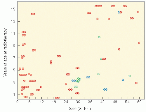
Figure 19.9 The relationship of radiation dosage and age in the production of slipped capital femoral plates (blue circles). The green circles are other reported cases. Refer to Fig. 2 of Ref. 84 from which data were obtained. Normal epiphyseal plates are represented by orange circles. (From Silverman CL, Thomas PR, McAlister WH, et al. Slipped femoral capital epiphysis in irradiated children: dose, volume, and age relationships. Int J Radiat Oncol Biol Phys. 1981;7:1357-1363, with permission).

Avascular Necrosis (Osteonecrosis). Avascular necrosis (osteonecrosis [ON]) is reported now with increased frequency following not only RT, but also following treatment with corticosteroid (Fig. 19.10). The rate of self-reported avascular necrosis was evaluated in the CCSS. Fifty-two cancer survivors reported ON in 78 joints, yielding 20-year cumulative incidence of 0.43% and a rate ratio (RR) of 6.2 (95% CI, 2.3-17.2) compared with sibling and 44% developed ON in a previous radiation field. Risk was greatest among survivors of stem-cell transplantation for leukemia. Nontransplantation patients with leukemia and bone sarcoma were at higher risk for ON. Older age at diagnosis, shorter elapsed time, older treatment era, exposure to dexamethasone (± prednisone), and gonadal and nongonadal radiation were independently associated with ON (86).

Even among patients treated with more contemporary therapy for ALL or NHL without RT, there is increasing information on the occurrence of ON, where the affected number is unclear, due to lack of systematic methods of detection. However, as many as 1 of 3 may be affected, likely reflecting the cumulative exposure to glucocorticosteroid therapy. The pathogenesis includes suppression of bone formation, expansion of the intramedullary lipocyte compartment and a direct effect on nutrient arteries. Children 10 years of age and older are at particular risk and the disorder is substantially more common in Caucasians than African Americans, and risk is higher in newer protocols with higher cumulative and more
continuous use of dexamethasone. Genetic predisposition may play a role. In an analysis from the Children’s Oncology Group, a PAI-1 polymorphism (rs6092) was associated with risk of osteonecrosis in univariate and multivariate analyses (adjusting for gender, age, and treatment arm) (87). Osteonecrosis is often multiarticular and bilateral, affecting weight-bearing joints predominantly (88, 89, 90, 91).


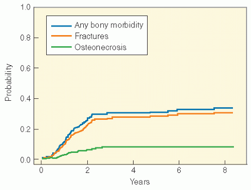
Figure 19.10 Cumulative incidence (CI) of bony morbidity for 176 children treated for ALL between 1987 and 1991. With 7.6 years of median follow-up, the overall 5-year CI (±SE) was 30% ± 4%. The 5-year CI of fracture was 28% ± 3%, and of osteonecrosis, 7% ± 2%. (From Strauss AJ, Su JT, Dalton VM, et al. Bony morbidity in children treated for acute lymphoblastic leukemia. J Clin Oncol. 2001;19:3066-3072, with permission.)


Other Skeletal Abnormalities

A variety of other skeletal abnormalities can be seen after irradiation. These include sternal deformity (hypoplasia, asymmetry, pectus excavatum, pectus carinatum) (92); hypoplasia of the iliac bones or lower ribs; cartilaginous exostoses; (93) osteochondromata (94); hypoplasia of the mandible; deformity of orbital, maxillary, nasal, or temporal bone (94,95); and lower extremity abnormalities such as acetabular dysplasia, coxa vara, hip dislocation, and leg shortening (96).

Table 19.6 reviews several musculoskeletal late effects with respect to causative treatments, signs and symptoms, screening and diagnostic tests, and management and intervention (41).

Osteopenia. Bone mineral density in childhood cancer survivors may be reduced, especially in children treated for ALL, or with HSCT, where it has been best studied. Risk factors include increased age at the time of exposure, estrogen deficiency, female sex, corticosteroid use and type, GH deficiency, and cranial radiation. Prevalence, chronicity, and severity are not consistent across studies, so the risk remains poorly defined (97, 98, 99, 100). Further research into the pathophysiology, the contribution of the various risk factors, the type and frequency of screening, the populations at highest risk, and interventions are clearly indicated.

Body Composition. Similarly of concern, but not yet rigorously studied, is abnormal body composition, which is reported in survivors of pediatric ALL. Oeffinger (101) evaluated obesity in 1764 ALL survivors followed in the CCSS and compared them with a cohort of 2565 siblings. The odds ratio for being obese was 2.6 for female survivors and 1.9 for male survivors who received dosages greater than 20 Gy. The highest risk was for girls treated at 0-4 years of age and with cranial radiation dosages greater than 20 Gy (Fig. 19.11). Risk of obesity was not higher among ALL survivors treated with chemotherapy alone or with cranial radiation dosages of 10-19 Gy. Similar findings were reported by Warner et al. (102), where body mass index Z-score, skinfold thickness, percentage fat by dual-energy x-ray absorptiometry, and ratio of central to peripheral fat was higher in girls treated for ALL than in siblings or patients treated for other malignancies. Reilly et al. (103) found higher obesity rates in survivors of childhood ALL, with higher risk in younger children and those thinner at the time of diagnosis, and associated with premature adiposity. However, there are also data that children treated for ALL in the absence of cranial RT are still at increased risk of obesity and hypertension, which may persist for years after therapy ends, as reported in a retrospective series by Chow (104). A follow-up study in the CCSS, where the use of cranial RT was common, also reveals continued risk of obesity among adult survivors (105). Increased body mass index is a component of the metabolic syndrome, which includes insulin resistance, hyperglycemia, hyperinsulinemia, hypertension, hyperlipidemia, and obesity. Long-term risk for cardiovascular disease from metabolic syndrome is of concern (102,104,106).


Cardiac

The functional and structural complexity of the heart places it at risk for a spectrum of RT and chemotherapy injuries that can occur. The radiation-associated sequelae include acute pericarditis during radiation (rare and associated with juxtapericardial cancer); delayed pericarditis, which can present abruptly or as chronic pericardial effusion; pancarditis, which includes pericardial and myocardial fibrosis with or without endocardial fibroelastosis (only after large dosages); myopathy in the absence of significant pericardial disease; coronary artery disease (uncommon), usually involving the left anterior descending artery; functionally valvular injury; and conduction defects (107, 108, 109). The hallmark of these injuries histologically is fibrosis in the interstitium with normal-appearing myocytes and capillary and arterial narrowing.

Several parameters must be considered in the evaluation of these radiation-associated injuries, including the relative weighting of the radiation portals and, therefore, the amount of radiation delivered to different depths of the heart; the presence of juxtapericardial tumor; the volume and specific areas of the heart irradiated; the total and fractional irradiation dosage; the presence of other risk factors in each patient such as age, weight, blood pressure, family history, lipoprotein levels, and habits such as smoking; and the use of specific chemotherapeutic agents.

The effects of thoracic RT are difficult to separate from those of anthracyclines because few children are exposed to thoracic RT in the absence of anthracyclines. The pathogenesis of injury differs, however, with radiation affecting primarily the fine vasculature of the heart and anthracyclines directly damaging myocytes. However, with current techniques and reduced dosages of RT, these effects are unlikely after

treatment for childhood cancer. Review of data from the CCSS shows significant cardiovascular morbidity across a number of childhood cancer survivor diagnostic groups, with the caveat that the data are based on self-report. Gurney et al (105). identified that 18% of childhood brain tumor survivors reported a heart or circulatory late effect. Risk was highest among those treated with surgery, RT, and chemotherapy compared to surgery and RT alone, suggesting a potential additive vascular injury from chemotherapy. Among ALL survivors in the CCSS cohort reporting a chronic medical condition, the risk of reporting a cardiac condition was nearly sevenfold higher compared to the siblings. No significant association was identified based on radiation exposure. A similar analysis among acute myeloid leukemia (AML) survivors in the cohort found the 20-year cumulative incidence of cardiac disease to be 4.7. Twenty-one percent of rhabdomyosarcoma survivors reported ≥1 cardiac sequelae compared to siblings. Among survivors of non-Hodgkin lymphoma (NHL), the standardized mortality ratio (SMR) for cardiac disease was 6.9 (110). A recent follow-up study of Wilms tumor survivors reported a cumulative risk of congestive heart failure of 4.4% at 20 years for those who received doxorubicin as part of their initial therapy, and 17.4% at 20 years where doxorubicin was received as part of therapy for relapsed disease. Risk factors for congestive heart failure in this cohort included female sex, lung irradiation with dosages 20 Gy or higher, left-sided abdominal irradiation, and doxorubicin dosage of 300 mg/m2 or more (111). Finally, cardiac complications after BMT may occur, with arrhythmias, pericarditis, and myopathies predominating. High-dose cyclophosphamide is clearly a causative agent. TBI is a secondary contributing factor.


Table 19.6 Evaluation of Patients at Risk for Late Effects: Musculoskeletal


Causative Treatment


Signs and Symptoms


Screening and Diagnostic Tests


Management and Intervention


Late Effects


Chemotherapy


Radiation


Surgery


Muscular hypoplasia


>20 Gy (growing child)


Younger children more sensitive


Muscle loss or resection


Asymmetry of muscle mass when compared with untreated area


Decreased range of motion Stiffness and pain in affected area (uncommon)


Careful comparison and measurement of irradiated and unirradiated areas


Range of motion


Prevention: good exercise program, range of motion, muscle strengthening


Spinal abnormalities: scoliosis, kyphosis, lordosis


For young children, RT to hemiabdomen or spine (especially hemivertebral)


Laminectomy


Back pain


Hip pain


Uneven shoulder height


Standing and sitting height at each visit and plot on chart (stadiometer), During puberty examine spine q3-6 months until growth is completed and then ql-2 years


Refer to orthopedist if any curvature is noted, especially during a period of rapid growth


Decreased sitting height


10 Gy (minimal effect)


>20 Gy (clinically notable defect)


Rib humps or flares


Deviation from vertical curve


Gait abnormalities


Spinal films for baseline during puberty, then PRN curvature (Lippman-Cobb technique to measure curvature)


Length discrepancy


>20 Gy


Lower back pain, limp, hip pain, discrepancy in muscle mass and length when compared with untreated extremity, scoliosis


Annual measurement of treated and untreated limb (completely undressed patient to ensure accurate measurements)


Radiograph baseline to assess remaining epiphyseal growth Radiographs annually during periods of rapid growth


Contralateral epiphysiodesis Limb-shortening procedures


Pathological fracture


>40 Gy


Biopsy


Pain, edema, ecchymosis


Baseline radiograph of treated area to assess bone integrity, then PRN


Prevention: consider limitation of activities (e.g., contact sports)


Surgical repair of fracture; may need internal fixation


Osteonecrosis


Steroids


40-50 Gy (more common in adults)


Pain in affected joint, limp


Radiograph, CT scan PRN


Symptomatic care


Joint replacement


Osteocartilaginous exostoses


RT


Painless lump or mass noted in the field of radiation


Radiograph for baseline and PRN with growth of lesion


Resection for cosmetic or functional reasons, counsel regarding 10% incidence of malignant degeneration


Osteopenia and osteoporosis


Steroids


>18 Gy cranial RT


Fractures, pain


Dual-energy x-ray ab: sorptiometry intervals of testing unclear Pediatric norms not well established. Best data are in adults


Calcium supplementation


Increase weight-bearing exercise


Refer to endocrinology for possible bis-phosphonate therapy


Slipped capitofemoral


High-dose steroids


>25 Gy (at young age)


Pain in effected hip, limp, abnormal gait


Radiograph baseline to assess integrity of the treated joints, then PRN


Refer to orthopedist for surgical intervention


RT, radiotherapy; PRN, as needed; CT, computed tomography.


From Constine LS, Hobbie W, Schwartz C. Facilitated assessment of chronic treatment by symptom and organ systems. In: Schwartz C, Hobbie W, Constine L, et al., eds. Survivors of Child and Adolescent Cancer: A Multi-Disciplinary Approach. Berlin: Springer-Verlag; 2005:17-34, with permission.


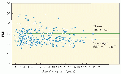
Figure 19.11 Scatterplot for unadjusted body mass index (BMI) by age at diagnosis of acute lymphoblastic leukemia for females treated with >20 Gy cranial radiotherapy. (From Oeffinger KC, Mertens AC, Sklar CA, et al. Obesity in adult survivors of childhood acute lymphoblastic leukemia: a report from the Childhood Cancer Survivor Study. J Clin Oncol. 2003;21:1359-1365, with permission.)


Pericarditis

Delayed acute pericarditis can be symptomatically occult or present suddenly with fever, dyspnea, pleuritic chest pain, friction rub, ST and T wave changes, and decreased QRS voltage. Up to 30% of patients treated for HL with a mean midplane heart dosage of 46 Gy are affected (112). With equally weighted anterior and posterior fields and the use of subcarinal blocking, the frequency decreases to 2.5% (113). The onset of delayed acute pericarditis averages 6 months, and 92% of effusions occur within 12 months. Although the effusion usually resolves in 1-10 months, it may persist for years. Pericardiectomy is a high-risk procedure in this setting because of the coexistence of other types of radiation-induced damage such as fibrosis of the myocardium and lung, coronary artery and valvular disease, impaired chest wall healing, and the patient’s general condition. However, Hancock et al. (114) noted a 4% (at 17 years) actuarial risk of pericardiectomy (occurring only in children treated with higher radiation dosages), and most patients improved after surgery.

Only gold members can continue reading. Log In or Register to continue

Stay updated, free articles. Join our Telegram channel

Jun 19, 2016 | Posted by in GENERAL RADIOLOGY | Comments Off on Late Effects of Cancer Treatment

Full access? Get Clinical Tree

Get Clinical Tree app for offline access