Second Primary Cancers


Second Primary Cancers


Smita Bhatia

Louis S. Constine


Significant improvements in therapeutic options and supportive care strategies have resulted in 5-year survival rates now approaching 65% for adult-onset cancers, and exceeding 80% in children (1). This improvement in survival has resulted in a growing population of cancer survivors. Currently in the United States, there are over 12 million cancer survivors. However, this survival is associated with significant morbidity. In fact, 60-70% of young adult survivors of childhood cancer are reported to develop at least one health-related complication (2); and second primary cancers (SPCs) are the most emotionally and physically devastating of these complications (Fig. 20.1).


SECOND PRIMARY CANCERS: THE SCOPE OF THE PROBLEM

SPCs are defined as histologically distinct malignancies developing at least 2 months after completion of the treatment for the primary malignancy. Several large epidemiologic studies have demonstrated that the SPC risk after adult-onset primary cancer is modest at best, ranging from no difference from the general population to a twofold greater incidence (3, 4, 5). A follow-up of a large cohort of childhood cancer survivors demonstrates that the cumulative incidence of second malignant neoplasms (SMNs) exceeds 3% at 20 years after diagnosis of a primary cancer in childhood, representing a sixfold increased risk for cancer survivors, compared to the general population (6). Furthermore, the absolute excess risk of SPCs after a first primary cancer diagnosed and treated during childhood is 1.9 per 1000 patient-years of follow-up of this cohort (6). Data from the Surveillance, Epidemiology, and End Results (SEER) program used to calculate the influence of SPCs on overall incidence trends in childhood cancer revealed higher annual incidence rates for all childhood cancers combined, specifically for acute lymphoblastic leukemia (ALL) and brain tumors, but not for other cancer types because of their rarity. However, excluding SPCs from the analysis had a negligible effect on the observed trends. These studies demonstrate that the relative risk of SPC in survivors of adult-onset cancer is modest at best, and that the high relative risk of SPC after a primary cancer in childhood does not translate into a high absolute risk. However, a recent report indicates that survivors of childhood cancer have a persistent excess risk of an SPC throughout their lives, accompanied by continuous changes in the risk of cancers at specific sites (7). This, combined with the fact that the morbidity and mortality associated with SPCs are substantial, necessitates a need to characterize this complication and identify associated risk factors.


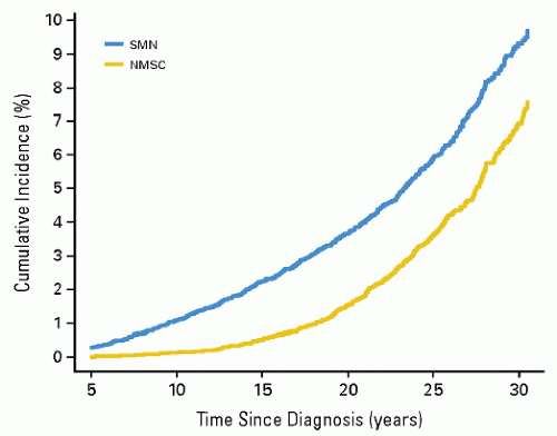
Figure 20.1 Cumulative incidence of second malignant neoplasms and nonmelanoma skin cancers in childhood cancer survivors. (From Meadows AT, Friedman DL, Neglia JP, et al. Second neoplasms in survivors of childhood cancer: findings from the Childhood Cancer Survivor Study cohort. J Clin Oncol. 2009;27:2356-2362, with permission).

The incidence and type of SPCs differ with the primary cancer diagnosis, type of therapy received, and presence of genetic conditions. Unique associations with specific therapeutic exposures have resulted in the classification of SPCs into two distinct groups: chemotherapy-related myelodysplasia and acute myeloid leukemia and radiation-related solid tumors. Characteristics of therapy-related myelodysplasia and acute myeloid leukemia include a short latency (<3 years from primary cancer diagnosis) and association with alkylating agents and/or topoisomerase II inhibitors (8). Solid SPCs have a strong and well-defined association with radiation, and are characterized by a latency that exceeds 8-10 years (8).

SPCs are a leading cause of nonrelapse late mortality, both in childhood cancer survivors (9), as well as in survivors of adult-onset cancer (10).


RISK FACTORS FOR SECOND CANCERS

Exposures to radiation therapy or to specific chemotherapeutic agents have been shown to increase the risk of SPCs in certain subsets of patients (6,11, 12, 13, 14). The types of SPC vary with the primary diagnosis, the type of therapy received, presence of genetic predisposition, and the time from initial
treatment. Follow-up of the Childhood Cancer Survivor Study demonstrates that, after adjusting for exposure to radiation, the risk of SPC was significantly associated with female sex (p < 0.001), childhood cancer diagnosed at a younger age (p < 0.001), initial cancer diagnosis of Hodgkin lymphoma (p < 0.001), and exposure to alkylating agents (p = 0.02) (6).


Primary Diagnosis

Hereditary retinoblastoma, Hodgkin lymphoma, and soft tissue sarcomas are over-represented among patients who develop SPCs relative to their incidence in the general population (6,14). This could result from an interaction between the genetic predisposition to develop cancer and the specific cancer therapies, as is clear in patients with hereditary retinoblastoma and familial soft tissue sarcoma (14,15). In individuals with other primary malignancies, such as Hodgkin lymphoma, it is not clear whether the primary diagnosis is an independent risk factor for the development of SPC or whether the specific therapy needed to treat the primary cancer is the major contributor to the development of the SPC. Some associations between first and second cancers are summarized in Table 20.1.


Table 20.1 Second Cancers and their Relationship with Primary Cancers


Second Cancers


Primary Cancers


Median Latency (Years)


Risk Factors


Breast cancer


Hodgkin lymphoma Bone tumors Soft tissue sarcomas Acute lymphoblastic leukemia Brain tumors Wilms tumors Non-Hodgkin lymphoma


15-20


Radiation Female sex


Brain tumors


Acute lymphoblastic leukemia Brain tumors Hodgkin lymphoma


9-10


Radiation Younger age


Myelodysplastic syndrome and acute myeloid leukemia


Acute lymphoblastic leukemia Hodgkin lymphoma Bone tumors


3-5


Topoisomerase II inhibitors Alkylating agents


Thyroid cancer


Acute lymphoblastic leukemia Hodgkin lymphoma Neuroblastoma Soft tissue sarcoma Bone tumors Non-Hodgkin leukemia


13-15


Radiation Younger age Female sex


Bone tumors


Retinoblastoma (heritable) Other bone tumors Ewing sarcoma Soft tissue sarcomas Acute lymphoblastic leukemia


9-10


Radiation Alkylating agents Splenectomy


Soft tissue sarcomas


Retinoblastoma (heritable) Soft tissue sarcoma Hodgkin lymphoma Wilms tumor Bone tumors Acute lymphoblastic leukemia


10-11


Radiation Younger age Anthracyclines


Host-Related Risk Factors


Age at Diagnosis and Treatment of Primary Cancer

Younger age at diagnosis of the primary cancer has been reported to be associated with a higher risk of radiation-associated SPCs (6,11,16). Conversely for secondary myelodysplasia and acute myeloid leukemia, which are strongly linked with specific chemotherapeutic agents, the risk increases with age at diagnosis and treatment of primary cancer (17,18).


Sex

Female sex is associated with a higher risk of SPCs, contributed to primarily by the excess occurrence of secondary breast and thyroid cancers among female cancer survivors (19). Moreover, some studies indicate a greater susceptibility of women to known carcinogens such as cigarette smoke (20). Possible mechanisms that underlie this greater susceptibility include greater activity of cytochrome P450 (CYP) enzymes, enhanced formation of DNA adducts, and the effects of hormones such as estrogen on tumor promotion (20).


Therapy-Related Risk Factors


Radiation

Although ionizing radiation is capable of causing most types of cancer, organs vary in their susceptibility. The risk is highest when exposure occurs at a younger age and increases with increasing dosages of radiation and with increasing follow-up from radiation (21). Radiation-associated SPCs have a long latency period and typically develop in or at the edge of the radiation field. Some well-established radiation-associated SPCs include breast cancer (after Hodgkin lymphoma), thyroid cancer (after Hodgkin lymphoma and ALL), lung cancer (after Hodgkin lymphoma), brain tumors (after other brain tumors and ALL), osteosarcoma (after retinoblastoma, Ewing sarcoma, and other soft tissue sarcomas), and nonmelanoma skin cancers (Table 20.1).

Several principles characterize radiation-induced SPCs (22, 23, 24):


  • A variety of histologic types of neoplasms can be induced by irradiation. These cancers are indistinguishable morphologically from naturally occurring cancers. The identification of a “radiation signature” in tumors would be important in evaluating SPC. There is evidence that radiation produces a different spectrum of mutations than other genotoxic agents. If these mutations could be discerned and characterized, then one could clearly identify radiation-induced tumors and understand, more completely, the radiation dose-response relationship for induced tumors. In this manner, molecular forensics may affect our understanding of attributable risk (25). Of interest is recent data showing that the risk of an SPC is proportional to the number of premalignant stem cells created, and surviving RT. This is related to cell killing by RT, cellular repopulation between RT fractions and after the last fraction, and the ratio of the proliferation rate of premalignant cells to the proliferation rate of normal cells (Fig. 20.2).


  • Low-linear energy transfer (low-LET) radiation (gamma ray, x-ray) is generally less efficient in inducing tumors than high-LET radiation. In a murine hepatocarcinogenesis model, for example, neutron irradiation produced a greater incidence of hepatomas than did gamma irradiation (26). Low-LET radiation appears to become less effective at carcinogenesis per centigray as the dosage falls, whereas high-LET radiation (neutrons, alpha particles) does not (27). With low-LET irradiation there is less tumor induction when the dosage is fractionated or administered at low dose rates; implying that repair of carcinogenic damage is occurring. With high-LET irradiation, the radiobiological effect is higher at low dosages. The carcinogenic effectiveness of high-LET radiation is not diminished and may be increased by dosage fractionation or protraction (28).


  • Comparison of the frequency of SPCs in different centers suggests that orthovoltage therapy is more likely to be carcinogenic than megavoltage therapy (24,29). This may be dose-related: By delivering a higher dosage to bone, orthovoltage irradiation may increase the risk of an SPC of bone. The higher risk may also be related to the long follow-up available for orthovoltage patients.


  • Although every tissue in the body is at risk for radiation-induced cancer, sensitivity varies according to the tissue. For example, thyroid gland and breast are sensitive to cancer induction after low radiation dosages; lymphoid tissue, lung, and liver are susceptible at moderate dosages, and bone at higher dosages. The relationship between dosage and response may vary according to the induced tumor. For example, the development of CNS tumors increases with increasing dose, whereas secondary thyroid carcinomas have a peak incidence after doses of 20-29 Gy but then decrease (Fig. 20.3A,B). Cancer risk from radiation may be given per unit dosage (gray, Gy) or per unit dosage equivalent (Sievert, Sv) where a “quality factor” (Q) is used to take account of the varying biological effectiveness of different radiations (e.g., for a gamma ray Q = 1, and for a neutron Q = 20). Therefore, dosage in Sv = dosage in Gy × Q. Detailed literature reviews indicate that the cancer mortality risk for the general population after whole body exposure is 1 × 10⁻⁴ to 4 × 10⁻⁴ per person-cGy. Pooled results of various partial body exposures give an estimated risk of 1 × 10⁻⁴ to 4 × 10⁻⁴ per person-cGy (4 × 10² Sv1). The risk based on incidence is about twice that for mortality (27,30). Leukemia data can be fit by a curvilinear dose-response model, whereas skin cancer appears to have a threshold dose-response function, and breast data fit a linear no-threshold model.


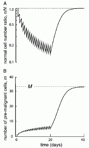
    Figure 20.2 Initiation/inactivation/proliferation model of carcinogenesis. Risk of SMN proportional to the number of premalignant stem cells created and surviving RT. Related to: (1) cellular killing, (2) cellular repopulation occurring between fractions and after the last fraction, and (3) the ratio of proliferation rate for premalignant cells to the proliferation rate of normal cells. (From Sachs RK, Brenner DJ. Solid tumor risks after high doses of ionizing radiation. Proc Natl Acad Sci U S A. 2005;102:13040-13045, with permission).


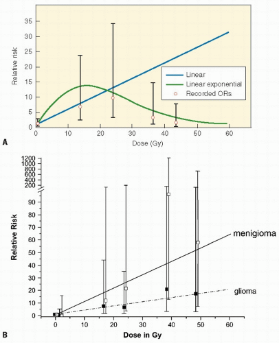
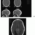
    Figure 20.3 A: Dose-response relations between radiotherapy dose and relative risk of second neoplasms. (From Meadows AT, Friedman DL, Neglia JP, et al. Second neoplasms in survivors of childhood cancer: findings from the Childhood Cancer Survivor Study cohort. J Clin Oncol. 2009;27: 2356-2362) B: Dose-risk for RT-induced CNS tumors from Childhood Cancer Survivor Study of 14,361 5-year survivors. (From Neglia JP, Robison LL, Stovall M, et al. New primary neoplasms of the central nervous system in survivors of childhood cancer: a report from the Childhood Cancer Survivor Study. J Natl Cancer Inst. 2006;98:1528-1537, with permission).


  • Radiation induces solid SMNs, within the radiation field and in a dose-dependent fashion (6,11,16,31, 32, 33, 34). A recent report assessed the relationship between the radiation dose received inside of the edges of radiation beams throughout the volume of tissue irradiated (thus the integral dose) and the likelihood of developing a secondary malignancy. In a cohort of 4401 patients who were 3-year survivors of all types of childhood cancer, a dose-response relationship was found between the overall risk of an SMN and the estimated integral dose (Table 20.2, Fig. 20.4) (35). As a corollary, data from Diallo et al. showed that a greater volume of tissues receives low or intermediate radiation doses in regions bordering the irradiated volume with modern multiplebeam radiation therapy administrations, including intensity modulated radiation therapy (IMRT) (36). However, in a report that modeled risk for children treated with IMRT versus conventional therapy, an increase was not seen (37). Additional data will be necessary to settle this issue.


  • SPCs may also be induced by agents other than radiation (chemotherapy, environmental exposures, hereditary disposition). The low-dose radiation dose-response curve could be influenced by these confounding factors to produce a result that is the sum of two independent rates or may be greater than simple addition would indicate. A variety of chemotherapeutic agents (to be discussed), especially alkylators, are known to be carcinogenic, and they may be additive or synergistic with radiation. Immunosuppressive agents clearly influence the propensity for tumor induction, as is seen in the setting of organ transplantation (38).


  • A large number of irradiated patients are necessary to calculate a radiation carcinogenesis risk with reasonable accuracy.


  • Latent periods vary according to the induced tumor. At least two patterns of latent periods for radiation-induced cancer have been described. The first, exemplified by the risk of leukemia in atomic bomb survivors, consists of an early wavelike pulse of increased risk followed by a gradual decline to baseline levels. The second, more typical of solid tumors, is an increase in relative risk of SPCs over many years, which remains constant over time thereafter. The latter pattern suggests that a multi-event pattern of carcinogenesis is involved: the initial event (radiation) is followed by promoting events (i.e., smoking, alcohol, or environmental exposures) over many years (39). Long latent periods complicate the study of radiation-induced cancer because the presence of other carcinogens or disease processes may not be well documented.


  • The duration of follow-up for any study population influences the frequency of tumors seen.


  • It is unclear whether the risk of tumor development after exposure is simply an absolute increase that is proportional
    to the radiation dosage or a relative increase that builds on an underlying spontaneous risk (greater for some patients) of developing a malignancy.


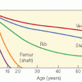
  • Age is a critical factor in determining radiation risk. In children, the most common SPCs occur in tissues undergoing rapid proliferation such as bone, thyroid, and breast. An actively proliferating tissue may be more susceptible to malignant transformation in any single cell because of the greater number of cell divisions. This might explain the higher frequency of secondary bone tumors in children than in adults (40). Childhood cancer survivors are likely to develop leukemias or sarcomas, whereas secondarily induced embryonal tumors are rare.


Table 20.2 Integral Dose as a Risk for Second Cancers


Integral Doseb


Integral Dose (kg)


Exposure


RRa


Exposure


RRa


0


0


1-46


2.42


0-4.3


2.42


47-179


2.64


4.3-7.5


2.78


179-1560


5.43


7.5-34.5


4.85


Chemotherapy


Alkylators


1.79


Intercalculating


1.81


agents


a Adjusted for age at Dx, sex, and dose.

b Total absorbed energy (joules), which depends on dose delivered, irradiated volume and its density, and estimated only for the volume inside the geometrical limits of the beam.


(Modified from Nguyen F, Rubino C, Guerin S, et al. Risk of a second malignant neoplasm after cancer in childhood treated with radiotherapy: correlation with the integral dose restricted to the irradiated fields. Int J Radiat Oncol Biol Phys. 2008;70:908-915, with permission).


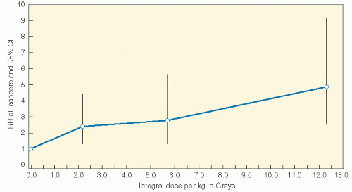
Figure 20.4 Integral dose and SMN risk in pediatric cancer survivors. (Modified from Nguyen F, Rubino C, Guerin S, et al. Risk of a second malignant neoplasm after cancer in childhood treated with radiotherapy: correlation with the integral dose restricted to the irradiated fields. Int J Radiat Oncol Biol Phys. 2008;70:908-915, with permission).


Chemotherapeutic Agents

Alkylating agents and topoisomerase II inhibitors have been shown to increase the risk of secondary myelodysplasia and acute myeloid leukemia (11,41,42). Alkylating agents have also been linked with bone tumors (43,44) and bladder cancer (45). Therapy-related myelodysplasia has been reported after treatment of Hodgkin lymphoma, non-Hodgkin lymphoma, ALL, and sarcomas (8,11,31,34,46,47), with the cumulative incidence approaching 2% at 15 years after therapy (11). The incidence of therapy-related myelodysplasia and acute myeloid leukemia typically peaks 4-6 years from diagnosis of the primary cancer and reaches a plateau after 15 years. The clinical observation that the risk of therapy-related leukemia does not extend beyond 15 years, despite an increasing risk of second neoplasms at other sites, indicates that the at-risk population of cells is no longer present (23). It may be that this period of time allows early pluripotent hematopoietic progenitors to undergo clonal extinction and be replaced from the compartment of resting stem cells by clonal succession. It is possible that stem cells in the resting phase of the cell cycle are relatively protected from the genotoxic effects of chemotherapy and radiation and that the excess risk of therapy-related leukemia would diminish as undamaged cells were recruited into active hematopoiesis. Therapy-related leukemia is associated with a very poor outcome, with an estimated 12-month survival of 10% (48).

Therapy-related myelodysplasia and acute myeloid leukemia is a clonal disorder characterized by distinct chromosomal changes (48, 49, 50). Two types are recognized by the WHO classification: alkylating agent-related type and topoisomerase II inhibitor-related type (51).


Alkylating Agent-Associated Acute Myeloid Leukemia and Myelodysplasia

Table 20.3 summarizes the incidence, risk factors, and outcomes among patients with therapy-related leukemia after alkylating agent exposure (11,41,52, 53, 54, 55, 56, 57, 58, 59). Alkylating agents associated with t-MDS/AML include cyclophosphamide,
ifosfamide, mechlorethamine, melphalan, busulfan, nitrosoureas, chlorambucil, dacarbazine (60), and platinum compounds (61). Mutagenicity is related to the ability of alkylating agents to form crosslinks and/or transfer alkyl groups to form DNA monoadducts. Alkylation results in inaccurate base pairing during replication and single- and double-strand breaks in the double helix as the alkylated bases are repaired. The risk of alkylating agent-related t-MDS/AML is dose-dependent, with a latency of 3-5 years after exposure (62); it is associated with abnormalities involving chromosomes 5 (− 5/del[5q]) and 7 (−7/del [7q]), and a high frequency of multidrug-resistance phenotype (63). The 5q31-33 region of the long arm of chromosome 5 contains at least nine genes involved in hematopoiesis. Defects in any of these genes could disrupt the balance between cell growth and differentiation and play a role in initiation and progression of leukemia. Complete or partial deletions of the long arm of chromosome 7 (7q— and -7) are nonrandom abnormalities observed in therapy-related leukemia.


Table 20.3 Characteristics of Therapy-Related Myelodysplasia or Acute Myeloid Leukemia After Treatment with Alkylating Agents and Topoisomerase II Inhibitors


Reference


Primary Cancer


Cohort Size


Cases of MDS and AML


Latency (Years)


Cumulative Probability (%) (Years)


Risk Factors


Percentage Alive


Associated with alkylating agents


53


HL


694


8


51.6 (16-148)


1.5 (20)


Chemotherapy Relapse


0


54


HL


1641


7


NA


0.8 (30)


NA


NA


55


HL


667


5


49 (31-125)


1.1 (15)


Alkylating agents


0


56


Childhood cancer


9170


19


NA


0.8 (20)


Increasing age


NA


HL, Ewing’s Alkylating agents Doxorubicin


11


HL


1380


24


48 (10-168)


2.8 (14)


Alkylating agents Older age


0


Associated with topoisomerase II inhibitors


57


ALL


734


21


39.5 (15-100)


3.8 (6)


Etoposide: weekly or twice weekly


14


58


ALL


205


10


32


5.9 (4)


Etoposide


50


59


NHL


38


5


21


18.4 (4)


Etoposide, twice weekly


60


MDS, myelodysplasia; AML, acute myeloid leukemia; HL, Hodgkin lymphoma; NA, not applicable; ALL, acute lymphoblastic leukemia; NHL, non-Hodgkin lymphoma.


Topoisomerase II Inhibitor-Associated Acute Myeloid Leukemia

Topoisomerase II catalyzes the relaxation of supercoiled DNA by covalently binding and transiently cleaving and religating both strands of the DNA helix. DNA topoisomerase II inhibitors (epipodophyllotoxins and anthracyclines) stabilize the enzyme-DNA covalent intermediate, decrease the religation rate and cause chromosomal breakage. These events initiate apoptosis, required for antineoplastic activity (64). Occasionally, repair of chromosomal damage results in chromosomal translocations, leading to leukemogenesis (65,66). Most of the translocations disrupt a breakpoint cluster region between exons 5 and 11 of the band 11q23 and fuse mixed lineage leukemia (MLL) with a partner gene (67,68). In vitro studies provide evidence that epipodophyllotoxins can directly induce MLL rearrangements in hematopoietic cells (69). Topoisomerase II inhibitor-related t-AML presents as overt leukemia often with a high blast count, rather than an initial myelodysplastic presentation (Table 20.4) (70) with a predominance of monocytic phenotypes (M4 and M5), after a latency of 6 months to 3 years; and is associated with balanced translocations involving chromosome bands 11q23 or 21q22. Other translocations include inv(18) (p13q22) or t(17,19)(q22;q12) (70,71).

Dosing schedule has been associated with epipodophyllotoxin-related secondary leukemia (57). Within the subgroups of patients who received epipodophyllotoxins (etoposide or teniposide) twice weekly or weekly, the cumulative risks were 12.3% and 12.4%, respectively. Of the remaining subgroups that included patients who received epipodophyllotoxins every 2 weeks, did not receive epipodophyllotoxins, or received them only during remission induction, the cumulative risk was 1.6%. After adjustment for the frequency of treatment, there was no apparent independent effect of the total dosage of epipodophyllotoxins in this study (57). Another large study from the Cancer Therapy Evaluation Program of the National Cancer Institute described the cumulative risks at 6 years for the development of secondary leukemia to be 3.3%, 0.7%, and
2.2% at the low (less than 1.5 g/m2), moderate (1.5-3.0 g/m2), and high (more than 3.0 g/m2) dosages of etoposide, respectively, again suggesting a lack of dose-response relationship. A recent study using a case-controlled design explored the dose-response relationship between epipodophyllotoxins and anthracyclines, and the risk of developing secondary leukemia. This study concluded that patients who received 1.2-6.0 g/m2 of epipodophyllotoxins or more than 170 mg/m2 of anthracyclines have a risk seven times higher than that of patients who received lower dosages or none of these drugs (72).


Table 20.4 Features of Chemotherapy-Induced Hematopoietic Malignancies


Property


Alkylating Agents


Epipodophyllotoxins


Median latency


4-6 years (range, 1-20 years)


1-3 years (range 0.5-4.5 years)


Presentation


Myelodysplasia


Abrupt, no preleukemia


Cytogenetic abnormalities


Loss of genetic material, often from chromosomes 5 and 7


Balanced translocations (often include 11q23)


Age


Typically older patients


Younger patients


CONTEMPORARY UNDERSTANDING OF CARCINOGENESIS


Genetic Predisposition

Some cancers tend to aggregate in families, with specific constellations of tumor types observed (e.g., adenocarcinomas or sarcomas) (15,73). Some inherited syndromes clearly predispose patients to the development of SPCs. Patients with the nevoid basal cell carcinoma syndrome who are irradiated for medulloblastoma develop skin cancers in the irradiated fields 6 months to 3 years later. Children with ataxia-telangiectasia may be more prone to irradiation-induced malignancies. Members of families with Li-Fraumeni syndrome have been reported to be at higher risk of multiple subsequent cancers than the general population (12). The risk was highest among survivors of childhood cancer, and the excess risk was mainly for cancers characteristic of Li-Fraumeni syndrome, such as brain tumors and sarcomas. Patients with soft tissue sarcomas who develop an SPC are more likely to have a family history of cancer than those without one (15). The tumor types occurring in excess in family members are similar to those observed as SPCs, such as cancers of the breast, bone, joint, or soft tissue, thus indicating that the risk of SPCs is associated with a familial predisposition. Genetic predisposition also plays a significant role in increasing the risk of SPCs among patients receiving radiation therapy for hereditary retinoblastoma, with the risk increasing with increasing dosages of radiation (74). These studies suggest that germ-line mutations in tumor suppressor genes might interact with therapeutic exposures to increase the risk of SPCs.

Literature clearly supports the role of chemotherapy and radiation in the development of SPCs. However, interindividual variability exists, suggesting a role for genetic variation in susceptibility to genotoxic exposures. The risk of SPCs could potentially be modified by mutations in high-penetrance genes that lead to serious genetic diseases, for example, Li-Fraumeni syndrome (75) and Fanconi anemia (76, 77, 78, 79). However, the attributable risk is expected to be very small because of their extremely low prevalence. The interindividual variability in risk of therapy-related SPCs is more likely related to common polymorphisms in low-penetrance genes that regulate the availability of active drug metabolite, or those responsible for DNA repair. Genetic variation contributes 20-95% of the variability in cytotoxic drug disposition. Polymorphisms in genes involved in drug metabolism and transport are relevant in determining disease-free survival and drug toxicity (80). Variation in DNA repair plays a role in susceptibility to de novo cancer (81, 82, 83, 84, 85) and likely modifies SPC risk after exposure to DNA-damaging agents, such as radiation and chemotherapy. Gene-environment interactions may magnify subtle functional differences resulting from genetic variations.


Drug-Metabolizing Enzyme

Metabolism of genotoxic agents occurs in two phases. Phase I involves activation of substrates into highly reactive electrophilic intermediates that can damage DNA—a reaction principally performed by the CYP family of enzymes. Phase II enzymes (conjugation) function to inactivate genotoxic substrates. The phase II proteins comprise the glutathione Stransferase (GST), NAD(P)H:quinone oxidoreductase-1 (NQO1), and others. The balance between the two sets of enzymes is critical to the cellular response to xenobiotics; for example, high activity of phase I enzyme and low activity of a phase II enzyme can result in DNA damage from the excess of harmful substrates.


DNA Repair

DNA repair mechanisms protect somatic cells from mutations in tumor suppressor genes and oncogenes that can lead to cancer initiation and progression. An individual’s DNA repair capacity appears to be genetically determined (86). A number of DNA repair genes contain polymorphic variants, resulting in large inter-individual variations in DNA repair capacity (87). Even subtle differences in an individual’s DNA repair capacity may be important in the presence of large external influences such as chemotherapy or radiotherapy. Mismatch repair (MMR) functions to correct mismatched DNA base pairs that arise as a result of misincorporation errors that have avoided polymerase proofreading during DNA replication (88). Approximately, 50% of t-MDS/AML patients have microsatellite instability, associated with methylation of the MMR family member MLH1 (89,90), low expression of MSH2 (91), or polymorphisms in MSH2 (92). RAD51 is one of the central proteins in the homologous repair (HR) pathway, functioning to bind to DNA and promote ATP-dependent homologous pairing and strand transfer reactions (93).
RAD51-G-135C polymorphism is significantly over-represented in patients with t-MDS/AML compared with controls (C allele: OR = 2.7) (94). Although XRCC3-Thr241Met was not associated with t-MDS/AML (OR = 1.4, 95%CI, 0.7-2.9), a synergistic effect resulting in an eightfold increased risk of t-MDS/AML (OR = 8.1, 95% CI, 2.2-29.7) was observed in the presence of XRCC3-241Met and RAD51-135C allele in patients with t-MDS/AML compared with controls (94). Base excision repair (BER) pathway corrects individually damaged bases occurring as a result of ionizing radiation and exogenous xenobiotic exposure. The XRCC1 protein plays a central role in the BER pathway and also in the repair of single strand breaks, by acting as a scaffold and recruiting other DNA repair proteins (86,95). The protein also has a BRCA1 C-terminus (BRCT) domain—a characteristic of proteins involved in DNA damage recognition and response. The presence of variant XRCC1-399Gln has been shown to be protective for t-MDS/AML (96) and basal cell carcinoma (97). Nucleotide excision repair (NER) removes structurally unrelated bulky damage induced by radiation and chemotherapy. The polymorphic Gln variant (ERCC2 Lys751Gln) of the NER pathway is associated with t-MDS/AML (98).

Only gold members can continue reading. Log In or Register to continue

Stay updated, free articles. Join our Telegram channel

Jun 19, 2016 | Posted by in GENERAL RADIOLOGY | Comments Off on Second Primary Cancers

Full access? Get Clinical Tree

Get Clinical Tree app for offline access