Soft Tissue Sarcomas Other Than Rhabdomyosarcoma; Desmoid Tumor


Soft Tissue Sarcomas Other Than Rhabdomyosarcoma; Desmoid Tumor


Kenneth B. Roberts

Edward C. Halperin


The word sarcoma is derived from the Attic Greek word σ&aacgr;ρκωµα or sarkoma meaning a fleshy excrescence (1). The term was used by Galan with particular reference to a nasal growth (2). An early English language dictionary from 1657 continued this definition of sarcoma as a “flesh growing in the nostrils like the proud flesh in a sore” (3). In modern usage, however, soft tissue sarcomas are defined as all malignant tumors of nonepithelial, extraskeletal tissues including the peripheral and autonomic nervous system but excluding the hematopoietic system, glia, and supporting tissues of specific organs and viscera. Soft tissue sarcomas constitute approximately 6.5-7% of childhood cancer. Within this 6.5-7%, approximately one half are rhabdomyosarcoma (4, 5, 6). Because the distribution of the types of other soft tissue sarcomas in children is distinctly different from that in adults, Spunt and Pappo have advocated the term “nonrhabdomyosarcoma soft tissue sarcoma” (NRSTS) (7). This group of soft tissue sarcomas constitutes 3-3.5% of childhood cancers (8), affecting approximately 500 children less than 20 years of age in the United States each year. NRSTS has not received the same focus of attention as rhabdomyosarcomas—though similar in incidence—perhaps because they have proved to be more resistant to conventional chemotherapy and radiotherapy (RT). While the surveillance, epidemiology and end results (SEER) database does not distinguish among rhabdomyosarcomas and NRSTSs, pediatric soft tissue sarcomas in the United States have been associated with improving survival over time. For patients less than 16 years of age diagnosed with a soft tissue sarcoma in 1975-1977, the adjusted 5-year survival was 61%. For those diagnosed in 1996-2004, there was a statistically significant improvement in 5-year survival at 74% (9,10). But to what degree this simply reflects improvements in rhabdomyosarcoma management is difficult to know. Moreover, very few patients with NRSTSs have been enrolled in cooperative group clinical trials. For instance in the United States prior to the 2006 Children’s Oncology Group (COG) ARST0332 trial, less than 200 patients had been enrolled in only three prospective clinical trials in the United States (11, 12, 13).

For most children with NRSTS, the origin or cause of the tumor is unknown. Some cases may be traced to prior radiation exposure (see Chapter 20), chemical exposure, iatrogenic or disease-caused immunosuppression, and neurofibromatosis, with the latter group having a 7-10% lifetime risk of developing malignant peripheral nerve sheath tumor (MPNST, historically called a neurofibrosarcoma or malignant schwannoma). The association of sarcomas with neurofibromatosis indicates that some sarcomas are associated with chromosomal deletions and translocations and the presence of abnormalities of tumor suppressor genes. Homozygous gene deletions occur in both the long and short arms of chromosome 17 in neurofibromatosis type 1. Candidate tumor suppressor genes include 17q11 (the NF tumor suppressor gene) and p53 (17p13) (4). Rhabdomyosarcoma and NRSTS also occur as part of the familial Li-Fraumeni syndrome. More generally in adult and pediatric soft tissue sarcomas, cytogenetic abnormalities are common with a mixture of simple translocations or point mutations giving rise to fusion genes or oncogenes and more complex genetic instability with a multiplicity of chromosome aberrations (14, 15, 16, 17, 18, 19, 20, 21, 22, 23, 24, 25, 26, 27, 28, 29, 30, 31) (Table 12.1). Gastrointestinal stromal tumors are of particular interest albeit rare in children since its associated c-kit oncogene is a successful therapeutic target with imatinib, a prototype tyrosine kinase inhibitor (32). While not yet tested in pediatric NRSTS, there are a variety of other molecular targets and associated agents under investigation in adult soft tissue sarcomas including platelet-derived growth factor receptor-A (sunitinib), Raf kinase (sorafenib), mTOR (rapamycin), vascular endothelial growth factor (bevacizumab), heat shock proteins, hedgehog, histone deacetylase, and nucleotide excision repair (31).


PATHOLOGY

The frequency of the different histologic subgroups of NRSTS of childhood varies between reporting institutions (Table 12.2). These differences may be attributable to variations in referral patterns and to the small numbers in each series. In addition, NRSTSs often are difficult for pathologists to classify, and there is wide intraobserver variation (33). In an M. D. Anderson Hospital series of sarcomas of the head and neck in children and adolescents, histologic diagnoses were changed in 22% of patients (34). Several other studies have assessed discrepancy rates between the original diagnosis of soft tissue tumors and the diagnosis made by expert reviewers when patients are referred to specialty centers for entry in therapeutic trials. About 5-10% of cases having the original diagnosis of sarcoma are revised to nonsarcoma, and for 16-32% of patients with a sarcoma, the histologic subtype is revised. Where grade was analyzed, there was disagreement in up to 40% of the cases (35). For example, malignant fibrous histiocytoma (MFH) was first described as a separate entity in the 1960s and by the 1970s was widely accepted by pathologists, identifying this entity as the most common soft tissue sarcoma subtype. Therefore during this time decades ago, the reported incidence of MFH in adults sharply increased while that of fibrosarcoma fell (36). Historically, MFH has been unusual in childhood. But with general advances
in pathologic categorization based on immunohistochemistry, ultrastructural studies, and cytogenetics, the diagnosis of MFH has been questioned as a distinct entity (37). At least in adults, this is no longer a common diagnosis, and when undifferentiated it is generally considered the same as a high-grade pleomorphic sarcoma. 
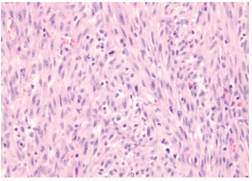
Figure 12.1 shows the histologic appearance of an MFH or pleomorphic sarcoma.


Table 12.1 Common Cytogenetic Changes in Nonrhabdomyosarcoma Soft Tissue Sarcomas


Histologic Type


Characteristic Cytogenetic Events


Genes Involved


Alveolar soft-part sarcoma


t(X;17)(p11;q25)


ASPSCR1-TFE3 (ASPL-TFE3) fusion


Aggressive fibromatosis (desmoid tumor)


Trisomies 8 and 20


APC inactivation


Deletion of 5q


Lipoma (typical)


12q15 rearrangement


HMGA2 (HMGIC) rearrangement


Well-differentiated liposarcoma


Ring form of chromosome 12


Myxoid/round-cell liposarcoma


t(12;16)(q13;p11)


FUS-DDIT3 (FUS-CHOP) fusion


t(12;22)(q13;q12)


EWSR1-DDIT3 (EWS-CHOP) fusion


Lipoblastoma


Rearrangement of 8q11-13


PLAG1 gene rearrangements: HAS2/PLAG1, COLIA2/PLAG1


Pleomorphic liposarcoma


Complex abnormalities


Malignant fibrous histiocytoma


Complex abnormalities


Myxoid malignant fibrous histiocytoma


Ring form of chromosome 12


Low-grade fibromyxoid sarcoma


t(7;16)(q34;p11)


FUS-BBF2H7 fusion


Leiomyoma (uterine)


t(12;14)(q15;q24) or deletion of 7q


HMGA2 (HMGIC) rearrangement


Leiomyoma (extrauterine)


Deletion of 1p


Leiomyosarcoma


Deletion of 1p


Other complex abnormalities


Monophasic synovial sarcoma


t(X;18)(p11;q11)


SS18-SSX1 (SYT-SSX1) or SS18-SSX2 (SYT-SSX2) fusion MYCN overexpression


Biphasic synovial sarcoma


t(X;18)(p11;q11)


SS18-SSX1 (SYT-SSX1) fusion MYCN overexpression


Benign schwannoma


Deletion of chromosome 22


NF2 inactivation


Malignant peripheral nerve sheath tumors, low-grade


None


Malignant peripheral nerve sheath tumors, high-grade


Complex abnormalities


Primitive neuroectodermal Tumor


t(11;22)(q24;q12)


EWSR1-FLI1 (EWS-FLI1) fusion


t(21;22)(q12;q12)


EWSR1-ERG (EWS-ERG) fusion


Desmoplastic small round-cell tumor


t(11;22)(p13;q12)


EWSR1-WT1 (EWS-WT1) fusion


Dermatofibrosarcoma protuberans


Ring form of chromosomes 17 and 22


COL1A1-PDGFB fusion


t(17;22)(q21;q13)


Endometrial stromal tumor


t(7;17)(p15;q21)


JAZF1-SUZ12 (JAZF1-JJAZ1)


Gastrointestinal stromal tumor


Monosomies 14 and 22


KIT of PDGFRA mutation


Deletion of 1p


Fibrosarcoma, infantile


t(12;15)(p13;q26)


ETV6-NTRK3 fusion


Trisomies 8, 11, 17, and 20


Extraskeletal myxoid chondrosarcoma


t(9;22)(q22;q12)


EWSR1-NR4A3 (EWS-NR4A3) fusion


t(9;17)(q22;q11)


TAF15-NR4A3 (TAF2N-NR4A3) fusion


Inflammatory myofibroblastic tumor


2p23 rearrangement


ALK fusion to TPM3, TPM4, clathrin, and other genes


Clear cell sarcoma


t(12;22)(q13;q12)


EWSR1-ATF1 (EWS-ATF1) fusion


Malignant rhabdoid tumor


Deletion of 22q


HSNF5 (INI1) deletion or mutation


Gene symbols are those provided in the Human Genome Nomenclature Database (http://www.genenames.org). Previous names of genes are given in parentheses.


Adapted from Skubitz KM, D’Adamo DR. Sarcoma. Mayo Clinic Proc. 2007;82(11):1409-32; and Spunt SL, Skapek SX, Coffin CM. Pediatric nonrhabdomyosarcoma soft tissue sarcomas. Oncologist. 2008;13(6):668-678.


Many of the childhood NRSTSs have characteristic cell types (Table 12.3). The WHO classification uses lines of differentiation to categorize tumors into adipocytic, fibroblastic/myofibroblastic, “so-called” fibrohistiocytic, smooth muscle, pericytic (perivascular), and vascular types, as well as tumors of uncertain differentiation (49). Of course, other sarcomas include the skeletal muscle
tumors/rhabdomyosarcomas and bone/cartilaginous tumors discussed elsewhere in this textbook.


Table 12.2 The Most Common Types of Childhood Nonrhabdomyosarcoma Soft Tissue Sarcoma in Recent Clinical Seriesa


Histology


St. Jude Children’s Research Hospitalb (6,83,223)


Harvard Joint Center for Radiation Therapyc (56)


Children’s Hospital of Philadelphia (224)


Baylor/Texas Children’s Hospital (8)


Children’s Hospital and Medical Center Seattled (4)


M. D. Anderson Hospital Head and Neck Sarcomas (34)


SEERe Data (66)


Italian Cooperative Study of Childhood Soft Tissue Sarcoma (147)


Pediatric Oncology Group (225)


Rambam Medical Center (68)


Mayo Clinic (48)


Instituto Nazionale Tumori Milan (226)


University of Iowa (162)


Primitive neuroectodermal tumor and extraosseous Ewing sarcoma and Askin tumor


32%


40%


36%


5%


Neurogenic sarcoma and neurofibrosarcoma


11%/18%


30%


38%


9%


15%


10%


2%


16%


3%


Synovial sarcoma


28%/15%


16%


10%


6%


10%


17%


10%


14%


42%


16%


36%


32%


24%


Alveolar soft part sarcoma


6%/12%


2%


2%


4%


2%


7%


5%


5%


Sarcoma not otherwise specified


14%


2%


30%


26%


15%


8%


Malignant fibrous histiocytoma


14%/12%


11%


15%


4%


4%


12%


5%


13%


1%


10%


Hemangiopericytoma


—/4%


2%


5%


2%


1%


4%


Liposarcoma


—/4%


2%


1%


2%


3%


8%


5%


Dermatofibrosarcoma protuberans


4%


5%


1%


2%


Fibrosarcoma


11%


9%


18%


4%


20%


17%


48%


14%


13%


33%


15%


5%


21%


Mixed mesenchymoma


2%


6%


Angiosarcoma and hemangiosarcoma


—/8%


2%


4%


3%


<2%


2%


6%


Epithelioid sarcoma


5%/8%


8%


2%


<2%


12%


8%


6%


Extrarenal rhabdoid sarcoma


—/8%


3%


1%


3%


Clear cell sarcoma


5%/8%


1%


8%


5%


2%


Leiomyosarcoma


1%


4%


5%


2%


7%


6%


Other


20%/16%


20%


4%


12%


2%


6%


13%


aIn each series, the most common histologic types are indicated by bold type.

b The number to the left of the slash is the number of surgically resectable patients; to the right are patients with metastatic disease.

c This series does not include children with metastatic disease.

d This series combines dermatofibrosarcoma protuberans with fibrosarcoma.

e Survival, epidemiology, and end results.


Figure 12.1 The histologic appearance of a malignant fibrous histiocytoma on hematoxylin and eosin staining, original magnification × 40. Many pathologists now categorize this lesion as a pleomorphic sarcoma. Note the spindle cell proliferation (long, narrow rodlike cells) with frequent mitoses, enlarged hyperchromatic and pleomorphic nuclei, and a storiform pattern of growth (fascicles of cells radiating from a central point). There is also some tendency toward a herringbone pattern in this example, characteristic of a fibrosarcoma.

Malignant peripheral nerve sheath tumor (MPNST) has also been known as neurofibrosarcoma, neurogenic sarcoma, malignant schwannoma, or malignant neurilemmoma. It is a malignant neoplasm that arises in a peripheral nerve sheath. In children, from one fifth to two thirds of cases of MPNST are associated with neurofibromatosis type 1 (NF1) (50, 51, 52). The morphology is characterized by fascicles of spindle cells with a herringbone or storiform pattern. One may observe evidence of schwannian differentiation (53). There can be areas of significant nuclear hyperchromatism and abundant mitotic figures (54). These tumors usually are positive for S-100, vimentin, and neuron-specific enolase. In about 2-16% of patients with NF1, nodular and plexiform neurofibromas transform to MPNSTs. NF1 is transmitted as an autosomal dominant with variable penetrance. However, almost 50% of cases are sporadic mutations (1). People with NF1 are 1.2 times more likely to have a malignant neoplasm listed on their death certificates than those who do not have the disease. These include MPNST, optic gliomas, and pheochromocytomas (55). NF1 is also called the von Recklinghausen disease. Friedrich Daniel von Recklinghausen (1833-1910) was born in Guterslah, Westphalia, Germany, and studied medicine at the Universities of Bonn, Würzburg, and Berlin. He was a professor in Konigsberg, Wurzburg, and Strasbourg. Also known for coining the term “hemochromatosis,” he published a monograph in 1882 that first described NF1 and the accompanying benign neurofibromas having a histologic appearance of disordered peripheral nerves and fibrosis.


Table 12.3 Cells of Origin of Nonrhabdomyosarcoma Soft Tissue Sarcoma of Childhood


Cell of Origin


Sarcoma


Fat


Liposarcoma


Blood and lymphatic vessels


Angiosarcoma, hemangiopericytoma


Fibroblasts


Fibrosarcoma, malignant fibrous histiocytoma


Smooth muscle


Leiomyosarcoma


Nervous tissue


Malignant peripheral nerve sheath tumor


Synovial


Synovial sarcoma


Chondrocytic


Extraskeletal chondrosarcoma


Epithelial


Epithelioid sarcoma


Melanocytes


Clear cell sarcoma of tendons and aponeuroses


Myofibroblasts


Myofibrosarcoma


Based on Pappo AS, Parham DM, Rao BN, et al. Soft tissue sarcomas in children. Semin Surg Oncol. 1999;16:121-143.


Biphasic synovial sarcoma, the more common type, also has spindle-shaped cells. These are mixed with oval and keratin-positive epithelial cells. Pseudoglandular spaces or slits and clefts mimic synovium (56). About one third of synovial sarcomas in children are the monophasic type (57). About two thirds of cases involve the lower extremity and one third the upper. Synovial sarcomas differ from other NRSTSs in that they have a significant risk of lymph node metastases (57,58). The monophasic type may have a better prognosis, but this is debatable (57,46). Synovial sarcomas commonly have a t(x;18)(p 11.2;q11.2) translocation that may result in two different fusion genes (SYT-SSX1 and SYT-SSX2). Controversy exists as to the prognostic importance of these fusion genes. Initial reports suggested that SYT-SSX1 fusion was associated with improved survival and less potential for metastasis (59,60). But in a contradictory small series, the SYT-SSX1 fusion was associated with a 42% 5-year metastasis-free survival, compared with 89% for the SYT-SSX2. The SYT-SSX1 fusion is also associated with a higher cell proliferation rate as assessed by Ki-67 staining (61). Yet another recent report suggests tumor grade rather than fusion gene subtype in synovial sarcoma is the more important prognostic factor (62).

Liposarcomas originate from primitive mesenchymal cells rather than from mature adipose tissue. Some have a myxoid appearance, whereas others resemble benign lipomas. The pleomorphic type may resemble fibroblastic, myoblastic, or synovial sarcoma. Angiosarcoma should always be considered high grade with an aggressive behavior, a high rate of local recurrence, a propensity to metastasize, and a poor prognosis (63).

Leiomyosarcoma originates from smooth muscle. The well-differentiated lesions usually have a centrally located blunt-ended nucleus (“cigar-shaped”).

Fibrosarcoma is an infiltrative, fibrous neoplasm composed of interlocking bundles of spindle cells (64,65). The tumor usually stains positive for vimentin. There are two clinically different
forms of fibrosarcoma in children. One is a lesion appearing in the first 5 years of life with a low rate of distant spread. This type of fibrosarcoma, called the congenital type, is generally treated by excision (4,33,
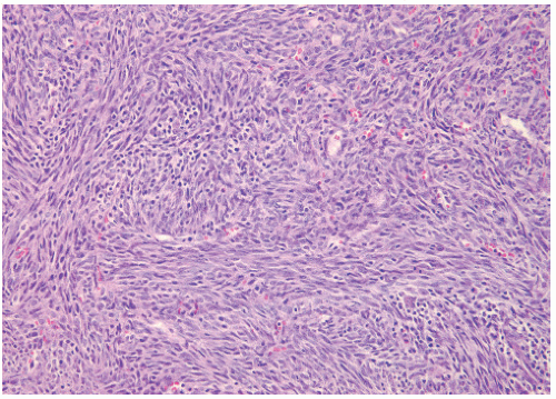
40). In children younger than 5 years of age this tumor may also be called infantile fibrosarcoma (Figure 12.2). The other occurs in children older than 5 years and has a more ominous prognosis with behavior similar to that of adult forms (65). This classic or adult-type fibrosarcoma is treated according to the principles outlined for other NRSTSs. Infantile fibrosarcoma has a rapid initial growth but generally indolent behavior. Local recurrence is common, metastases are rare, spontaneous regressions have been reported, and RT is rarely used. Infantile and adult fibrosarcomas are histologically identical. There is no routine microscopic, immunohistochemical, or ultrastructural way of distinguishing the two clinical types (64). Low-grade fibromyxoid sarcoma is an indolent tumor that rarely occurs in children. It consists of spindle and stellate cells with uniform nuclei arranged in a whorled pattern with alternating areas of fibrous and myxoid stroma (66). True malignant fibrous histiocytomas are pleomorphic sarcomas as noted above, often characterized by a whorled growth pattern. They are thought to arise from histiocytic cells acting as facultative fibroblasts. Hemangiopericytomas arise from pericytes, the modified smooth muscle cell with contractile function located on the internal surface of venous capillaries and postcapillary venules (67). The malignant mesenchymoma has two or more cell types, any of which, taken by itself, might be considered a malignant neoplasm (68,69); however, the term is loosing favor as a clinicopathologic entity as such sarcomas can usually be classified in other ways (70). Ectomesenchymomas are felt to be of neural crest origin with multidirectional differentiation exhibiting combinations of neuroblastoma, ganglioglioma, schwannoma, embyronal rhabdomyosarcoma, benign melanocytic proliferation, and bone or cartilage elements. These rare tumors may have a dominant rhabdomyosarcomatous element with clinically similar response to chemotherapy and RT (71) (Figure 12.3).


Figure 12.2 Example of microscopic pathology of an infantile fibrosarcoma, original magnification ×20. This is another fibrous spindle-cell proliferation as in the example of an MFH. While histologically aggressive in appearance, when this fibrosarcoma occurs in the first several years of life there tends to be a benign biologic behavior with little potential for metastatic spread. Local excision is the primary management. In unresectable cases chemotherapy can be used to promote regression and subsequent surgery.

There are several childhood NRSTSs that have a characteristic microscopic picture, but the cell of origin is uncertain. The epithelioid sarcoma is a tumor of the subcutaneous tissue, tendons, and fascia, usually of the upper extremity, including the hand. There is a nodular arrangement of plump, polygonal to round, epithelioid cells interspersed with spindle-shaped cells (5,72). Central degeneration or necrosis is often present. The tumor tends to spread within fascial planes or aponeuroses and may grow along the neurovascular bundle and encroach on large vessels or nerves. Regional lymph node metastasis may occur in association with high-grade tumors and tumors larger than 5 cm (73). The tumor generally stains positive for keratin.

Alveolar soft part sarcomas have a characteristic crystalline material seen with periodic acid-Schiff (PAS) stain (74,42). The tumor tests positive for vimentin on immunohistochemistry. The tumor cells typically have an organoid or nestlike arrangement. Vascular invasion is always seen. Of 11 children with alveolar soft part sarcoma seen at St. Jude Children’s Research Hospital in a 32-year period, 6 had localized disease and 5 had unresectable or metastatic disease. Cytogenetic studies indicate that 17q25 abnormalities are common (42). In 19 patients seen in Italy, 4 had metastatic disease at presentation. The 5-year survival for all 19 patients was 80% (75). A slow doubling time of the tumor may explain its very late occurrences (sometimes more than 10 years) (37,45).

Extraskeletal (or extraosseous) Ewing sarcoma (EOES) and peripheral primitive neuroectodermal tumor (PNET) are characterized by cohesive, uniform, small hyperchromatic cells in a fibrous background. Dense clumping of chromatin, mitotic figures, and rosette formation are typical of PNET. On immunohistochemistry analysis, EOES are generally positive for vimentin and HBA-17. PNET is generally positive for neuron-specific enolase and other neuron-related markers such as S-100 protein, neurofilament, or HNK-1. Both PNET and EOES are associated with a particular chromosome translocation t(11;22)(q24;q12). The progenitor cell for these two small, round, blue cell NRSTSs is not established. They may arise from neural crest, primordial germ cells, or perhaps mesenchymal stem cells (4,76). When PNET or EOES arises in the thoracic cavity it is called the Askin tumor.

Desmoplastic small, round, blue cell tumor is a rare intraperitoneal malignancy occurring predominantly in adolescent boys. It is characterized by a reciprocal translocation t(11;12)(p13;q12) associated with the EWS-WT1 gene fusion transcript. Cells often stain positive for desmin, keratin, and neuron-specific enolase. The predominant pattern of relapse is intraperitoneal (77). Patients generally are treated with surgical debulking, alkylator chemotherapy, and whole abdomen irradiation or intra-abdominal P-32. The relapse-free survival rate is approximately 20% (78,79) (50,51). PET scanning may be useful in follow-up for early diagnosis of recurrence (80). 
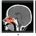
Figure 12.4 depicts a rare instance of this disease occurring outside the peritoneum.

Clear cell sarcoma is characterized by ovoid or polygonal cells with abundant clear cytoplasm, indistinct borders, large nucleoli, and abundant intracytoplasmic glycogen. Immunocytochemistry often is positive for S-100 protein, neuron-specific enolase, and melanocyte-associated antigen HMB-45. A specific chromosomal translocation t(12;22)(q13;q12) involving the DNA transcription factors ATF-1 on chromosome 12 and the EWS gene on chromosome 22 has been described in 60-75% of clear cell sarcoma cases (81,82).

A fair proportion of NRSTSs show no cellular differentiation. These are called undifferentiated sarcomas or sarcomas not otherwise specified.


Figure 12.3 This 10-month-old boy developed a 2.5 cm superficial mass in the right supraclavicular fossa. After ultrasound showed this to be a solid lesion, a marginal excision was performed in February 2003. An ectomesenchymoma was diagnosed having immature cartilage separated by bands of collagen in a myxoid background with cellular areas having primitive ovoid and spindle cells. The dominant cellular pattern had features of an embryonal rhabdomyosarcoma. Surgical margins were positive. There was no evidence of metastasis in an adjacent lymph node or on CT imaging. As a wider excision was not possible due to tumor abutting clavicle, lung apex, and carotid artery, postoperative RT was indicated. Along with VAC chemotherapy as per rhabdomyosarcoma management, the patient received 3060 cGy in 17 fractions using a four-field technique (AP, PA, RAO, LAO fields) facilitated by daily general anesthesia with IV propofol. Shown is the radiation treatment plan and digital reconstructed radiographs (DRR) of three of the fields. The treatment plan minimized the volume of distal clavicle, spine, and shoulder girdle being treated to full dose. In last follow-up at age 7 years without evidence of disease, there is negligible boney asymmetry seen on chest x-rays correlating with normal physical examination. Panel A: radiation treatment plan. Panel B: RAO DRR. Panel C: AP DRR. Panel D: LAO DRR. Panel E: follow-up CXRS in 2003, 2006, and 2009.


Figure 12.4 This 12-year-old female presented with a subcutaneous mass over the left shoulder that was marginally excised. Pathology showed a desmoplastic small, round-cell tumor with a classic t(11;12) translocation on cytogenetics. This case represents an unusual extra-abdominal presentation of this rare sarcoma. MRI scan showed contrast enhancement in the surgical bed (upper panel). Re-excision, axillary node dissection, bone marrow biopsy, and CT scans were negative for other sites of disease. The patient was treated with an Ewing sarcoma type chemotherapy protocol with involved-field RT to 45 Gy in 25 fractions (middle panel of axial, coronal, and sagital CT images with superimposed radiaton dose distribution). Despite local control of the primary tumor, the patient unfortunately developed progressive pulmonary metastases shortly after completing vincristine/doxorubicin/cyclophosphamide alternating with ifosfamide/etoposide chemotherapy. (See bottom panel of four serial CT images showing progression of lung metastasis over 6 months.) Despite surgical excisions of lung nodules and salvage chemotherapy, the patient ultimately succumbed to her disease.


PRESENTATION, WORKUP, AND STAGING

Most NRSTSs present as a painless swelling but may also present with signs and symptoms of vascular compression, neurologic impairment from nerve compression, or bowel dysfunction when tumors arise from the retroperitoneum.

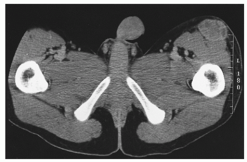
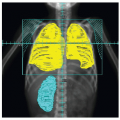
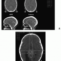
The radiographic workup begins with the plain radiograph. One looks for evidence of soft tissue mass, calcification, and destruction of adjacent bone. Radionuclide bone scanning is used to assess metastatic bone involvement, activity in bone adjacent to the tumor, and active vascular activity in the tumor itself. Arteriography is advocated for its delineation of the tumor’s blood supply, a matter of concern to the surgeon or to the direct infusional chemotherapist. Computed tomography (CT) and magnetic resonance imaging (MRI) are the essential studies for clear definition of tumor extent, patterns of infiltration, evaluation of adjacent bone, and planning surgical and radiotherapeutic approaches. MRI often shows a larger area of involvement than does CT (Fig. 12.5). The evaluation for distant metastasis focuses on the most common site, the lungs, with chest radiograph and thoracic CT scanning (51,52,56,83, 84, 
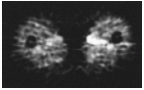
85). Metabolic scanning techniques, such as thallium scans and positron emission tomography (PET), are being used with increasing frequency. PET measures glucose utilization rate in sarcomas and may be used to assess lesion grade and to monitor neoadjuvant therapeutic response (Fig. 12.6) (86,87).

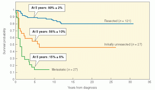
NRSTS in childhood may be staged by one of two systems. A few investigators still use the rhabdomyosarcoma grouping system, although their number is shrinking (see Table 11.2). This system is convenient and relies on surgical resectability as an important prognostic factor. There is no doubt that tumor size and resectability are important in predicting outcome in NRSTS of children. In fact, a retrospective analysis of NRSTS from the St. Jude Children’s Research Hospital has crudely identified three distinct risk groups: (1) grossly resected nonmetastatic disease (89% 5-year survival); (2) initially unresectable, nonmetastatic disease (56% 5-year survival); and (3) metastatic disease (15% 5-year survival) (38,88,89) (Fig. 12.7). Adverse risk factors include metastatic disease, tumor size >5 cm, high grade, positive surgical margins, intra-abdominal primary tumor site, and omission of postoperative RT in localized disease. Regarding tumor size as a prognostic factor in sarcoma patients (including rhabdomyosarcoma and NRSTS), a recent report suggests that there should be an adjustment for overall patient size (90). The authors noted, for example, that the mortality risk associated with a patient with a body surface area of 1.75 m2 and a 5-cm tumor was the same as for a 0.6 m2 child with a 2.8-cm tumor.


Figure 12.5 This 17-year-old white boy presented with a 7-month history of a mass, noted on self-examination, of the left thigh. The mass eventually grew to 4 cm in size. Magnetic resonance imaging demonstrated a mass without the involvement of bone. Positron emission tomography showed increased metabolic activity. On pathology, the tumor was “hypercellular highly vascularized neoplastic proliferation of generally small round to oval nuclei with evenly distributed chromatin and small nucleoli. Immunohistochemical stains are positive for vimentin, negative for leukocyte antigen; most consistent with extra skeletal Ewing’s sarcoma/primitive neuroectodermal tumor.” The patient was treated with tumor excision, involved-field irradiation, and systemic chemotherapy.


Figure 12.6 A positron emission tomography scan showing increased metabolic activity in a limb involved by primitive neuroectodermal tumor.

Histologic tumor grade, however, is of considerable importance and is not directly considered in the rhabdomyosarcoma grouping system (33,91, 92, 93). Sarcoma grade assessment incorporates pleomorphism, spontaneous necrosis, and number of typical and atypical mitoses per 10 high-power fields. Building on the first tumor grading system (squamous cell carcinoma of the lip) in 1920, Broders and colleagues at the Mayo Clinic published a grading system for sarcomas in 1939 based on the degree of mitosis, tumor giant cells, and fibrous stroma (94,95). In 1969, the American Joint Commission (AJC) on Cancer Staging described a staging system for soft tissue sarcoma that uses grade, tumor size, nodal involvement, and presence of metastases as the determinants of stage (Table 12.4). First validated by Russell et al. in 1977, the AJC system incorporates in the sarcoma staging system both TMN and tumor grade components (96). Grade is determined by evaluation of the degree of cellularity, cellular anaplasia or pleomorphism, mitotic activity, expansive or infiltrative growth, and necrosis (72). Frequency of distant metastases increases and the survival probability decreases with increasing size of the primary tumor (92). About 15% of patients have metastatic disease at presentation (4).

Histologic grading is an important way to predict the outcome of NRSTS. Moreover, as treatment decisions may often hinge on grading, the clinician should be aware of the criteria and uncertainties inherent in pathologic interpretation. There are various strategies for grading. One system, developed by the Pediatric Oncology Group (POG), labeled grade 1 as tumor that has little propensity for malignancy. Grade 2 tumors are those with fewer than 5 mitoses per 10 high-powered fields or less than 15% geographic necrosis, and grade 3 tumors are those known to be
clinically aggressive by virtue of histologic diagnosis and with more than 4 mitoses per 10 high-powered fields or more than 15% geographic necrosis (97) (Table 12.5). One review of this POG grading system found a 73% mortality in grade 3 lesions and 15% mortality in grade 1 and 2 tumors (98).


Figure 12.7 Risk stratification of nonrhabdomyosarcoma soft tissue sarcomas. (From Spunt SL, Hill DA, Motosue AM, et al. Clinical features and outcome of initially unresected nonmetastatic pediatric nonrhabdomyosarcoma soft tissue sarcoma. J Clin Oncol. 2002;20:3225-3235, with permission.)


Table 12.4 The 2010 American Joint Committee on Cancer Staging System for Sarcoma of Soft Tissuea


T: Primary tumor


Tx


Primary tumor cannot be assessed


T0


No evidence of primary tumor


T1


Tumor less than or equal to 5 cm in greatest dimension


T1a


Superficial tumorb


T1b


Deep tumorb


T2


Tumor more than 5 cm in greatest dimension


T2a


Superficial tumorb


T2b


Deep tumorb


N: Regional lymph nodes


Nx


Regional lymph nodes cannot be assessed


N0


No regional lymph node metastasis


N1


Regional lymph nodes metastasis


M: Distant metastasis


MX


Distant metastasis cannot be assessed


M0


No distant metastasis


M1


Distant metastasis


G: Histopathologic gradingc


Gx


Grade cannot be assessed


G1


Well differentiated


G2


Moderately differentiated


G3


Poorly differentiated


Staged


Stage IA


T1a/b N0 M0 G1 or Gx


Stage IB


T2a/b N0 M0 G1 or Gx


Stage IIA


T1a/b N0 M0 G2 or G3


Stage IIB


T2a/b N0 M0 G2


Stage III


T2a/b N1 M0 G2


T2a/b N0 M0 G3


Stage IV


M1


aThis staging system does not apply to Kaposi sarcoma, gastrointestinal stromal tumors (which has its own classification), fibromatosis (desmoids tumor), infantile fibrosarcoma, or sarcomas arising in dura mater or brain, parenchymal organs, and hollow viscera. Staging system now includes dermatofibrosarcoma (previously excluded), angiosarcoma, and extraskeletal Ewing sarcoma.

b Superficial, above superficial fascia without invasion of the fascia; Deep, located either beneath the superficial fascia or superficial to the fascia with invasion of or through the fascia. Retroperitoneal, mediastinal, and pelvic sarcomas are classified as deep.

c AJCC now recommends a three-tier system. The FNCLCC (French) system is the preferred grading system.

d New in seventh edition of AJCC Cancer Staging Manual: There is now a staging distinction between G2 and G3, at least for tumors >5 cm.; N1 is now Stage III instead of Stage IV; Stage I is divided into IA and IB. Stage II is divided into IIA and IIB; clinical versus pathologic staging is designated cT versus pT; deep or superficial location of tumor is no longer a factor in overall stage.


From Edge SB, Byrd DR, Compton CC, et al., eds. AJCC Cancer Staging Manual, 7th ed. New York: Springer, 2010, with permission from AJCC.


Table 12.5 POG Grading System for Pediatric Nonrhabdomyosarcoma Soft Tissue Sarcomas


Grade


1


Liposarcoma: myxoid and well differentiated


Deep-seated dermatofibrosarcoma protuberans


Fibrosarcoma: well differentiated or infantile (<5 yr)


Hemangiopericytoma: well differentiated or infantile (<5 yr)


Well-differentiated malignant peripheral nerve sheath tumor


Angiomatoid malignant fibrous histiocytoma


2


All NRSTSs not in grades 1 or 3; <15% of tumor shows geographic necrosis, or mitotic index is <5 mitoses/10 high-power fields


3


Fibrosarcoma with >15% of tumor with geographic necrosis or mitotic index >5 mitoses/10 high-power fields


Liposarcoma: pleomorphic, round cell


Mesenchymal chondrosarcoma


Extraskeletal osteosarcoma


Malignant triton tumor


Alveolar soft part sarcoma


Based on Pappo AS, Parham DM, Rao BN, et al. Soft tissue sarcomas in children. Semin Surg Oncol. 1999;16:121-143.


The more common sarcoma grading systems that one is more likely to encounter in clinical usage are the National Cancer Institute (NCI) system and the French Federation of Cancer Centres (FNCLCC) system despite being largely based on adult cases (99). Both are a three-tiered grading system. The aforementioned POG system is a variation of the NCI classification scheme. In the NCI system (100), certain types of sarcomas are deemed low or high grade based on the histological subtype. A typical liposarcoma is low grade while epithelial or synovial sarcomas are always high grade, for instance. For those subtypes that are not automatically allocated to a grade, the degree of necrosis is the most important factor distinguishing intermediate versus high grade. Minor pathologic factors include the degree of mitosis, pleomorphism, cellularity, and stromal matrix. Necrosis as a prognostic marker is categorized by being minimal (0-15%), moderate (15-30%), or marked (>30%). Thus, when degree of necrosis is key to determining grade, the significant cutoff value is 15%. A grade 2 soft tissue sarcoma (e.g., a fibrosarcoma or an MFH) has up to 15% necrosis, while grade 3 disease is simply determined by greater than 15% necrosis.

The FNCLCC has similarities to the NCI system, but has a point scoring system based on tumor histology, necrosis, and mitosis that has a certain clinical appeal in its reproducibility and simplicity (101). The main criticism, however, is that some histologic subtypes (e.g., MFH, alveolar soft parts sarcoma) do not have attributes that recapitulate normal tissues and cannot be easily scored in terms of differentiation (Tables 12.6 and 12.7). Worldwide, the French system predominates.

Comparisons between the NCI and FNCLCC system show up to one third of cases have grading discrepancies, but the latter system may be a slightly better determinant of prognosis, albeit not for every NRSTS subtype (99,102). The current COG protocol on NRSTS has central pathology review; one of its aims will be to directly compare the POG and the FNCLCC grading systems for pediatric sarcomas. To date, the WHO or other pathology associations have not endorsed any particular grading schema.


Table 12.6 Tumor Differentiation Scores of Sarcoma in the French Federation of Cancer Centres System of Grading Soft Tissue Sarcomas


Diagnosis


Differentiation Score


Well-differentiated liposarcoma


1


Well-differentiated fibrosarcoma


1


Well-differentiated MPNT


1


Well-differentiated leiomyosarcoma


1


Well-differentiated chondrosarcoma


1


Myxoid liposarcoma


2


Conventional fibrosarcoma


2


Conventional MPNT


2


Well-differentiated malignant hemangiopericytoma


2


Myxoid MFH


2


Typical storiform/pleomorphic MFH


2


Conventional leiomyosarcoma


2


Myxoid chondrosarcoma


2


Conventional angiosarcoma


2


Round-cell liposarcoma


3


Pleomorphic liposarcoma


3


Dedifferentiated liposarcoma


3


Poorly differentiated fibrosarcoma


3


Epithelioid malignant schwannoma


3


Poorly differentiated MPNT


3


Malignant triton tumor


3


Conventional malignant hemangiopericytoma


3


Giant cell and inflammatory MFH


3


Poorly differentiated/epithelioid/pleomorphic leiomyosarcoma


3


Synovial sarcoma


3


Rhabdomyosarcoma


3


Mesenchymal chondrosarcoma


3


Poorly differentiated/epithelioid angiosarcoma


3


Extraskeletal osteosarcoma


3


Extraskeletal Ewing sarcoma/PNET


3


Alveolar soft part sarcoma


3


Malignant rhabdoid tumour


3


Clear cell sarcoma


3


Undifferentiated sarcoma


3


MPNT, Malignant peripheral nerve tumor; PNET, primitive neuroectodermal tumour; MFH, malignant fibrous histiocytoma.


Modified from Guillou L, Coindre JM, Bonichon F, et al. J Clin Oncol. 1997;15:350, with permission.


Table 12.7 French Federation of Cancer Centres System of Grading Soft Tissue Sarcomas


Parameter


Criterion


Tumor differentiation


Score = 1


Sarcoma histologically very similar to normal adult mesenchymal tissue


2


Sarcoma for defined histological subtype (e.g., myxoid MFH)


3


Sarcoma of uncertain type, embryonal and undifferentiated sarcomas


Mitosis count


Score = 1


0-9/10 HPF


2


10-19/10 HPF


3


≥20/10 HPF


Microscopic tumor necrosis


Score = 0


No necrosis


1


≤50% tumor necrosis


2


>50% tumor necrosis


Histological grade


Grade 1


Total score 2 or 3


2


Total score 4 or 5


3


Total score 6, 7, or 8


MFH, malignant fibrous histiocytoma; HPF, high-power field.


Modified from Guillou L, Coindre JM, Bonichon F, et al. J Clin Oncol. 1997;15;350, with permission.


While the clinician may rely on sarcoma grading to make treatment decisions, one must be aware of the degree of uncertainty in pathologic diagnosis. This problem has a bigger dimension as the pathologist is increasingly forced to evaluate smaller biopsy specimens rather than the whole tumor as newer treatment algorithms involve preoperative RT or chemotherapy. With smaller specimens, there is an increasing danger of sampling errors as necrosis, mitotic activity, differentiation, pleomorphism, and other grading aspects have geographical variability within a particular tumor. Despite advances in molecular biology, identification of cytogenetic or particular DNA or RNA lesions has not yet supplanted tumor grading to aid clinical decisions for NRSTS.


SELECTION OF THERAPY


Surgery

Every biopsy should be planned to be consistent with the treatment plan. The two techniques for the biopsy are either via needle or open incisional. Needle biopsy techniques include fine-needle aspiration (of variable accuracy and dependent on the experience of the cytopathologist) or core needle biopsy (4). The incision for an open incisional biopsy should be the minimum that is technically feasible and homeostasis should be secure. Incisions on the extremity should be in the long axis (85). Muscle compartments should not be crossed; one does not want to contaminate adjacent areas with tumor. The biopsy should be placed so that the entire surgical tract will be removed at the time of the definitive operation.

Complete surgical excision is the mainstay of therapy. NRSTS may infiltrate widely. Sarcomas tend to expand and infiltrate adjoining tissue spaces, producing a pseudocapsule made up of compressed normal tissue intermingled with microscopic extensions of the tumor. A system for assessing the adequacy of surgical margins in sarcoma surgery was described by Enneking et al. (103). An intralesional surgical margin is through tumor, with gross or microscopic contamination. A marginal resection is through the reactive or inflammatory zone. A wide excision is through normal tissue outside the inflammatory zone. A radical excision is outside the anatomic compartment containing the tumor. A wide excision or amputation often is needed to obtain the microscopic-free margin needed for control. Clinical experience, primarily in adults, has shown that the local failure rate for simple excision of malignant soft tissue sarcomas is 60-90% (51,52,56). This failure rate falls to 18-30% when simple excision is replaced by radical resection, radical compartmental resection, or amputation above the proximal joint (6,33,85,104, 105, 106, 107). In a pediatric series, the 5-year survival rate was higher for complete tumor excision than for partial excision (45). For low-grade lesions, wide excision with negative margins may be curative as the sole treatment (8,105). Surgical margins of at least 1.0 cm around gross tumor is preferred for optimal local control (108). In cases of positive margin or unanticipated finding of NRSTS on marginal excision, a re-excision should be strongly considered (109).

There are some patients for whom limb-sparing treatments may be considered (106,107,110, 111, 112). Limb-sparing surgery removes a soft tissue sarcoma while preserving the extremity with a satisfactory functional and cosmetic result. To achieve results comparable to those of radical procedures, most limb-sparing procedures involve the planned use of preoperative or postoperative external beam irradiation, brachytherapy, or intra-arterial or systemic chemotherapy. But regardless of adjuvant RT or chemotherapy, resection with negative surgical margins is critical for optimal local control (113). In the presence of planned RT, resection margins may be relatively tight within millimeters, but if surgery alone is planned margins greater than 1.0 cm are likely required along with an en bloc resection beyond any tumor pseudocapsule (114). For tumors abutting bone, stripping off the periosteum en bloc is usually sufficient in the absence of any direct bone invasion; however, the addition of RT in thigh sarcomas may entail a higher risk for later development of femoral bone fractures (115). Other investigators have observed an increased risk of bone fractures as simply a function of high radiation dose delivery and not necessarily related to periosteal resection (116,117). Sarcomas that involve neurovascular bundles are challenging clinical problems, in which surgical and adjuvant radiotherapeutic management must be individualized regarding the sacrifice of blood supply or nerves.

Limb-sparing is clearly not appropriate for some patients. These patients include young children who may deal better with an amputation than with the limb-length discrepancy that may occur with limb-sparing procedures, those with extremity lesions
where it is not possible to acquire adequate surgical margins and where RT may produce major long-term complications, those with lesions that involve major vessels or nerves where resection will severely compromise function, and those in whom a fracture has resulted from tumor and the limb is useless and painful (104,110). Simply put, in the skeletally immature patient, due consideration must be given to the possibility that inappropriate or incorrectly administered RT may produce a stiff, painful, shortened, or disfigured extremity and engender a risk of second malignant neoplasm. Therefore, in some situations amputation may be preferred.

The role of regional node dissection in NRSTS is evolving. Overall, the incidence of nodal involvement is about 4%, ranging from close to 0% for grade 1 to 12% for grade 3 (6,58). It is reasonable to biopsy enlarged regional nodes in high-grade primary tumors. Certain histologic subtypes are more prone to lymphatic spread, including epithelial and synovial sarcomas, while others are not. Kayton has published a small series using sentinel node biopsies in pediatric tumors suggesting its utility in NRSTSs that are at risk for lymphatic spread (118).

Surgery also plays a role in managing NRSTS in the treatment of pulmonary metastases (119,120). With proper patient selection, long-term survival is possible for those undergoing removal of pulmonary metastases. Preoperative evaluation with chest radiograph and thoracic CT scanning is performed to determine the number and location of the metastases. Tumor may be resected via a median sternotomy or lateral thoracotomy, generally with double-lumen endotracheal anesthesia. Two studies in the literature, both of which included adult and pediatric patients, evaluated the important prognostic factors predicting survival after pulmonary metastasectomy. Higher survival rates appear to be associated with complete resection of all metastases, the presence of few lesions (i.e., 1-3), and a long disease-free interval before the development of metastases. Patients rendered free of disease in the series of Jablons et al. (121) had a median survival of 26.8 months. A similar group in the series of Casson et al. (122) had a median survival of 28 months. It is only fair to note that there are no randomized series in the literature comparing surgery with observation or with chemotherapy without resection. Therefore, it cannot be proven that the apparent prolongation of survival after surgery is attributable to the surgical therapy as opposed to good tumor biology (121). As an alternative to surgical resection of oligometastatic disease, there is an evolving experience with so-called body radiosurgery (123). In essence, high-dose ablative and hypofractionated conformal RT is administered. With some of these radiosurgical techniques, the surgeon may be asked to assist with the placement of radio-opaque fiducial markers into the tumor mass to facilitate precise radiation dose delivery with minimal normal tissue exposures.

Only gold members can continue reading. Log In or Register to continue

Stay updated, free articles. Join our Telegram channel

Jun 19, 2016 | Posted by in GENERAL RADIOLOGY | Comments Off on Soft Tissue Sarcomas Other Than Rhabdomyosarcoma; Desmoid Tumor

Full access? Get Clinical Tree

Get Clinical Tree app for offline access