CHAPTER 46 Endolymphatic Sac Tumor
Epidemiology
This is a slow-growing tumor that arises from cells lining the endolymphatic sac. Most of the endolymphatic sac tumors (ELSTs) are sporadic. There is an association of ELST and von Hippel-Lindau (VHL) syndrome with the incidence of ELST, documented by magnetic resonance imaging (MRI), of 11% in patients with VHL. Patients with bilateral tumors are presumed to have VHL disease.
Clinical Features
Almost all patients present with hearing loss. Patients may also have symptoms of facial nerve palsy (60%), pulsatile tinnitus (50%), or even vertigo (20%).
Pathology
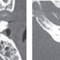
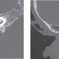
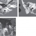
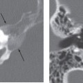
Macroscopically the tumors are reddish blue. These slow-growing tumors arising from the lining of the endolymphatic sac have a variable histopathologic appearance. Complex interdigitating papillary processes infiltrate surrounding connective tissue and bone. The papillary processes are generally embedded in sheets of dense fibrous tissue with recent and previous hemorrhage, cholesterol clefts, and scattered inflammatory cells. The papillary processes are lined with a single layer of low columnar to cuboidal epithelial cells resembling cells lining the endolymphatic sac. The cells have a small centrally placed ovoid nuclus with deeply eosinophilic homogeneous cytoplasm. There is no nuclear pleomorphism, and mitotic figures are absent. Tumor infiltration forms residual bone fragments that are seen as intratumoral calcifications on CT.
Treatment
The treatment of choice is complete surgical resection. There is no current role for radiation therapy or chemotherapy.
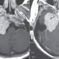
Imaging Findings
CT
Related posts:

Stay updated, free articles. Join our Telegram channel

Full access? Get Clinical Tree


