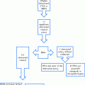
Grade
Type of occlusion
0
No occlusion
1
MCA occlusion (M3)
ACA occlusion (A2 or distal segments)
1 BA and/or VA branch occlusion
2
MCA occlusion (M2)
ACA occlusion (A1 and A2)
≥2 BA and/or VA branch occlusion
3
MCA occlusion (M1)
3a
Lenticulostriate arteries spared and/or leptomeningeal collaterals visualized
3b
No sparing of lenticulostriate arteries and no leptomeningeal collaterals visualized
4
ICA occlusion (collaterals present)
BA occlusion (partial filling, direct or via collaterals)
4a
Collaterals fill MCA
Anterograde filling
4b
Collaterals fill ACA
Retrograde filling
5
ICA occlusion (no collaterals)
BA occlusion (complete)
12.
During the procedure, the interventionalist should be constantly balancing the possible benefits and harms of pursuing recanalization depending on the elapsed time and the amount of lytics and devices used.
13.
Finally, the use of contrast should also be taken into account. Specially when administered through the micro catheter distal to the occlusion, contrast seems to increase the odds of hemorrhagic transformation (HT).
9 Post-Procedure Management and Follow-up Protocol
At the end of the procedure, the patient should be transferred to a Stroke Unit or Neurointensive Care Unit where strict control of blood pressure and glycemia are required. Successive NIHSS examinations may detect neurological deterioration indicating complications of the treatment. We also deploy TCD follow-up assessments of vessel patency to detect hyperemic reperfusion, re-occlusion, or impaired vasomotor reactivity—findings helpful to control blood pressure, decide on the need for an additional intervention, and ventilator management.
At 24 h from symptom onset, or if clinical deterioration occurs anytime, a repeat or new neuroimaging examination should be performed. This will allow determination of the infarct volume and possible complications such as HT or brain edema. Initiation of antithrombotic treatment for secondary prevention should be delayed until this second image is obtained, except in cases where a stent was deployed in which early use of anti-platelets is required to avoid in-stent reocclusion.
10 Intracranial Angioplasty and Stenting
In the last few years, development of intracranial stents is continuously increasing for the treatment of intracranial stenosis as well as acute occlusions. Several clinical trials are evaluating their efficacy in long-term outcome of intracranial atherosclerotic disease and the utility of retrievable stents for acute reperfusion. The indications about when an intracranial stenosis should be stented (i.e., after first event or after recurrence) are still to be defined.
Treatment of acute intracranial occlusions or severe symptomatic intracranial stenosis with angioplasty (with or without stenting) shows promising results. Angioplasty without stenting is probably associated with a high risk of re-stenosis or reocclusion.
11 Complications of Endovascular Treatment
The complications of the endovascular treatment of stroke can be related to the stroke itself or the vascular manipulation.
Stroke Complications
(a)
Symptomatic intracranial hemorrhage (sICH): it is probably the most feared complication of reperfusion treatment of acute ischemic stroke. Some hemorrhagic transformations (HTs) are asymptomatic, and may even be related to recanalization. These HTs do not imply a worse clinical evolution and some authors even associate them with a better outcome. On the other hand, sICH is associated with high mortality and worse clinical outcome. If sICH has occurred (confirmed by neuroimaging and clinical evaluation), fresh frozen plasma or coagulation factors can be administered. Poor outcomes are generally expected despite this treatment since sICH often occurs with most severe strokes at baseline. Probably, a very strict control of blood pressure may be the best way of preventing sICH.