Energy-Based Ablation of Other Liver Lesions


Energy-Based Ablation of Other Liver Lesions


Luigi Solbiati


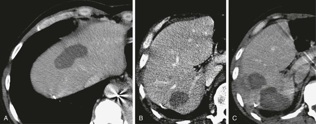
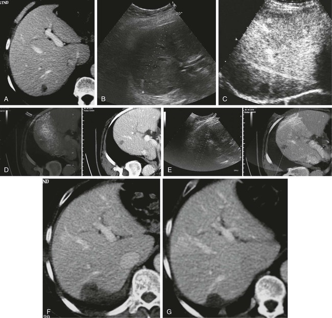
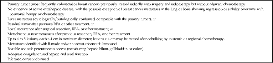
Metastatic liver disease is a very common issue in oncology practice. Currently, multiple treatment options are available, including hepatic resection, chemoembolization, intraarterial and systemic chemotherapy, cryotherapy, laser therapy, and radiofrequency ablation (RFA).1,2


Over the last few years, advances in diagnostic imaging modalities such as contrast-enhanced ultrasound, single- and multidetector helical computed tomography (CT), and magnetic resonance imaging (MRI) with hepatobiliary and reticuloendothelial-specific contrast agents have allowed early detection and accurate quantification of liver metastatic involvement.310 As a result, correct selection of patients for different treatment options is usually possible.


When feasible, surgical resection of hepatic metastases is the accepted standard therapeutic approach in patients with colorectal cancer and offers potential for cure in selected patients with other primary tumors.2,1128


RFA remains the most commonly used method of thermal ablation. Radiofrequency waves operate in the 30 kHz to 1 MHz range. Microwave ablation is also being performed with increasing popularity. Microwaves operate in the 1000 to 3000 MHz range. Microwave ablation can produce heat at a much faster rate than RFA, yielding shorter ablation times. In addition, the relatively rapid heat production with microwaves may minimize the impact of heat loss when lesions are in close proximity to large vessels. Microwave ablation is now being used frequently within the liver but may have particular advantages within the lung and bone, owing to relatively poor electrical conductivity within those tissues.


High-intensity focused ultrasound (HIFU) is yet another method of creating tumoricidal heat production. In the United States, HIFU is currently approved for treatment of uterine fibroids. Areas of investigation include prostate cancer, liver cancer, and brain cancers. The HIFU unit is coupled to and guided with MRI. Irreversible electroporation (IRE) is a method of rapidly increasing the sizes of pores within cell membranes, causing instantaneous cell death. Cell death is not based on thermal heating. IRE can interfere with the conduction system of the heart and is often performed with electrocardiographic gating.


Percutaneous (or laparoscopic) RFA is an established therapeutic option for liver metastases that may obviate the need for major surgery and result in prolonged survival and chance for cure. Extensive operator experience and technical advances provide larger coagulation volumes and therefore allow safe and effective treatment of medium and occasionally even large metastases. Results of RFA in terms of global and disease-free survival currently approach those reported for surgical metastasectomy. In published series, this technique has demonstrated significant advantages:


Indications


Colorectal Metastases


Patients with new metastases or local recurrence after previous hepatic resection, patients with multiple bilobar liver metastases, and those refusing or ineligible for surgery because of general health reasons are all candidates for RFA. Recently increased awareness among referring oncologists, satisfactory long-term survival reported in the scientific literature, and minimal invasiveness of RFA versus surgery have contributed to the widely accepted status of RFA as a valid therapeutic option for local treatment of patients with limited metastatic liver disease.


RFA is applicable to patients with one to five metachronous liver metastases measuring up to 3.5 to 4 cm in largest diameter (but preferably not > 3 cm) from previous radically treated colorectal cancer in whom surgery cannot be performed either because it is contraindicated or because it was simply refused.


Some patients with large lesions or more than five nodules may undergo RFA after successful tumor debulking by means of chemotherapy (neoadjuvant).


Noncolorectal Metastases


In our experience, RFA is a useful treatment in patients with metastases from previously treated malignancies, provided local control of liver disease may be beneficial from an oncologic perspective in terms of improved survival or quality of life. At our institution, patients with liver metastases from neuroendocrine, gastric, pancreatic, renal, pulmonary, uterine, or ovarian cancer and melanoma have been successfully treated with RFA.


Under these conditions, RFA has to be considered a less invasive therapeutic alternative to surgery. Indications for RFA of liver metastases from other primary cancers may follow those of surgical resection. Hepatectomy for liver metastases appears to be favorable in patients with gynecologic, gastric, and testicular primary tumors, with survival rates higher than 20%; unfortunately, no definite selection criteria have been reported in the literature.22,26,27


Particular considerations are reserved for patients with neuroendocrine tumors, which commonly metastasize to the liver. The frequency of metastatic disease, the hormone production, and the protracted clinical course make metastases from neuroendocrine tumors responsive to multiple therapeutic modalities, including surgical resection, hepatic artery embolization, cryotherapy, and RFA. Specific indications for RFA include patients who need intraoperative ablation as an adjunct to resection, who have limited hepatic disease but are not operative candidates, or who are inoperable and unresponsive to embolization treatments or experience recurrence after resection.


RFA can be applied to liver metastases from breast cancer when the liver is the only location of metastases, when extrahepatic metastases are demonstrated to be stable or in regression as a result of other treatment modalities, or when liver involvement is limited (one to five lesions, each 4 cm or less in maximum diameter) and systemic chemotherapy has been partially or completely unsuccessful.


Indications for ablation of hepatic metastases are summarized in Table 142-1.


Contraindications


Percutaneous RFA is a minimally invasive procedure, so there are few absolute contraindications to its use. Exclusion criteria include the presence of severe coagulopathy, renal or liver failure, portal vein neoplastic thrombosis, and obstructive jaundice. Active extrahepatic disease is a contraindication to RFA, with the exception of bone or lung metastases in breast cancer patients whose disease is responding or is unchanged with systemic chemotherapy.30


Contraindications to general anesthesia can be considered relative because the procedure can be performed under conscious sedation if necessary. Conscious sedation is not applicable if extended breath-holding is necessary during the procedure, depending on the position of the lesion within the liver.


Caution has to be exercised to not damage adjacent structures. Liver lesions adjacent to the hepatic hilum, gallbladder, stomach, and colon are potential candidates for ablation but require precise and careful planning. Adjacent vascular structures do not represent an obstacle alone; high blood flow in major hepatic vessels allows prompt dissipation of the warming effect secondary to RFA (heat sink effect), but the biliary system is vulnerable, especially if harboring bacteria. Proximity of the gastric wall or bowel loops to the area of treatment may be a critical safety issue when selecting candidates for treatment. Perforation of bowel loops by heating may occur many hours after ablation and could be clinically misleading in that it is usually less painful than perforation secondary to inflammatory diseases.


Relatively simple practical measures such as patient positioning, selection of a safe path to the target, and intraperitoneal injection of 500 mL of dextrose (to create “artificial ascites”) can provide adequate distance to avoid injury to critical structures.


Equipment


In most centers, thermal ablation of liver metastases is performed with radiofrequency energy (using both cooled-tip and multihooked needles), but cryoprobes can also be used. Microwaves are under investigation as an ablative modality and will soon become available commercially.


Single or cluster (three needles mounted on one handle) 16-gauge internally cooled electrodes are connected to a 200-W, 480-kHz RF generator system (Radionics, Burlington, Mass.). Ablation is impedance guided with an automated pulsed-RF algorithm. The choice of electrodes is based on the size of the target nodule. For lesions smaller than 2 cm, single insertion of a 3-cm exposed-tip single electrode is sufficient. Lesions 2 to 3 cm in size can be treated by single insertion of a cluster electrode or multiple insertions of a 3-cm exposed-tip electrode. Lesions exceeding 3 cm can be treated with only one to two insertions of a cluster electrode and one to two single-electrode insertions.43,44


The applied energy is variable and usually reaches 1600 to 1800 mA for single electrodes and 1800 to 2000 mA for cluster electrodes.45,46 Each application of energy lasts 8 to 12 minutes, and total procedure time ranges from 12 to 15 minutes for small solitary lesions to 45 to 60 minutes for large or multiple ablations.4749


As an alternative, four to eight hooked 14-gauge electrodes (RITA-AngioDynamics, Mountain View, Calif.; Boston Scientific, Natick, Mass.) connected to 100- to 200-W generators can be used. Temperature monitoring for each hook is available. Each application of energy lasts 10 to 15 minutes. For treatment of large lesions, 60- or 90-degree rotations of the electrode and multiple insertions are required, with a total treatment time of 50 to 60 minutes.


Cooled-tip and multihooked electrodes can achieve comparable volumes of necrosis. In general, ablation with cooled-tip electrodes is faster and technically easier, whereas multihooked electrodes allow accurate real-time monitoring of temperature at the periphery of treated targets. Because of the unfeasibility of real-time monitoring of the exact location of each hook, particularly with respect to “risky” anatomic structures, use of multihooked electrodes is technically more challenging than that of cooled-tip needles.


Technique


Technical Aspects


Patient enrollment in the treatment process requires evaluation by an anesthesiologist. Laboratory tests include a complete blood cell count (CBC), coagulation screen, urea, electrolytes, liver function tests, and tumor markers.


International guidelines recommend a dedicated operating room in which general anesthesia, endotracheal intubation, and mechanical ventilation can be optimally performed if needed. Standard surgical asepsis rules must be strictly observed by the operating team. Patients receive antibiotic prophylaxis (i.e., ceftriaxone) before treatment and antiemetic and analgesic drugs as needed postoperatively.


In our department, RFA sessions are carried out in a dedicated operating room. Conscious sedation is used in most patients. General anesthesia with endotracheal intubation and mechanical ventilation may be applied only for the treatment of lesions adjacent to the Glisson capsule (usually painful) or for risky anatomic structures.


RFA can be guided either by sonography or by fluoroscopy with CT. Sonography is used in most centers because of some favorable advantages: real-time control, low cost, and no use of ionizing radiation. CT guidance is used when sonographic targeting is not possible, but procedure time is significantly increased.


When sonography is used as the guidance modality, contrast-enhanced ultrasound is particularly useful for pretreatment planning, targeting of lesions undetectable in basal studies, and immediate evaluation of the early results of treatment.


B-mode and color/power Doppler ultrasound is unreliable in assessing the size and completeness of induced coagulation necrosis at the end of the application of energy. Furthermore, additional repositioning of the electrode is usually made difficult by the hyperechogenic “cloud” appearing around the distal probe. Therefore, we routinely perform contrast-enhanced ultrasound at the presumed end of the treatment session to enable rapid assessment of the extent of tissue ablation and detect viable tumor requiring additional immediate treatment.50


Keeping a “safety peripheral margin,” as in surgical treatment of tumor lesions, is crucial in the long-term outcome. A minimum margin of 0.5 cm is necessary; enlarging the target area by a 1-cm margin when feasible is highly recommended (Fig. 142-1).


Real-time ultrasound/CT fusion imaging technology allows targeting of lesions visible only with CT (Fig. 142-2), real-time calculation of the volume to be treated before treatment is undertaken, and guidance of further electrode insertion into the same target, which is often completely obscured for sonography by the gas formed during RFA.


In our experience, patients are hospitalized for 24 hours after treatment, with discharge following documentation of necrosis and exclusion of complications via a contrast-enhanced CT scan.


Dec 23, 2015 | Posted by in INTERVENTIONAL RADIOLOGY | Comments Off on Energy-Based Ablation of Other Liver Lesions

Full access? Get Clinical Tree

Get Clinical Tree app for offline access