Fig. 9.1
Epidemic cerebrospinal meningitis. (a, b) Transverse CT scanning demonstrates obviously enlarged cerebroventricular system including the four ventricles and communicating hydrocephalus. (c) Transverse FLAIR sequence demonstrates patches of high signals at the right posterior part of medulla oblongata. (d) Patches of high signals surrounding the triangular area in the right lateral ventricles and suppurative ependymitis (pointed by arrows) (Reprint with permission from vandebeek D, et al. Arch Neurol, 2007, 64(9): 1350)
Case Study 3
A male patient aged 17 years, with complaints of fever, nausea, and headache. The laboratory tests defined the diagnosis of epidemic cerebral meningitis.
For case detail and figures, please refer to Kastenbauer S, et al. Arch Neurol, 2001, 58(5): 806.
9.7.2 Pulmonary Infection
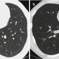
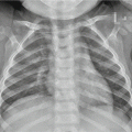
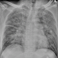
By chest X-ray, the demonstrations are not specific, including patches of shadows as bronchopneumonia and lobar infiltration which are commonly found in the inferior lobe and the right middle lobe, with accompanying pleural effusion in about 20 % of the cases.
9.8 Basis for the Diagnosis
9.8.1 Diagnosis of Epidemic Cerebral Meningitis
The suspected cases are commonly found in winters and springs and in epidemic areas, with a history of close contacts to patients with epidemic cerebral meningitis. The suspected cases also have symptoms of sudden high fever, nausea, vomiting, deteriorating headaches, petechiae and ecchymosis on skin and/or mucosa, irritation, delirium, coma, and convulsions, with accompanying signs of meningeal irritation. The early diagnosis should be made based on the cerebrospinal fluid examination and serum immunological essay.
The cases with imaging demonstrations of enlarged supratentorial ventricles, subdural effusion, and abnormal enhancement of meninges should be suspected as having epidemic cerebral meningitis.
9.8.2 Diagnosis of Epidemic Cerebral Meningitis-Related Complications
9.8.2.1 Subdural Effusion
After treatments for meningitis, the cases with persistent fever or recurrent fever, repeated convulsions, persistent protrusion of anterior fontanel, or recurrent protrusion of anterior fontanel after treatment should be suspected as having secondary subdural effusion. By subdural puncture, the discharged slightly yellowish fluid exceeds 2 ml. Its protein quantification is usually above 0.04 g/dl.
The diagnostic imaging demonstrates stripes of fluid signal in the peripheral brain, thickened meninges, and obvious abnormal enhancement by enhanced imaging.
9.8.2.2 Hydrocephalus
It has clinical manifestations of intracranial hypertension. By diagnostic imaging, the demonstrations include symmetrically enlarged supratentorial ventricles.
Stay updated, free articles. Join our Telegram channel

Full access? Get Clinical Tree


