Fig. 19.1
Pneumonic plague. (a) Chest X-ray demonstrates clear pulmonary markings in both lungs in nontreatment group of African green monkeys before their infection of Yersinia pestis; (b) chest X-ray demonstrates flakes of high-density shadows in the left lung field and in the right lower lung at day 5 after infection of Yersinia pestis; (c) chest X-ray demonstrates clear pulmonary markings in both lungs in the treatment group of African green monkeys before their infection of Yersinia pestis; (d) with medication of lenofloxacin immediately after the onset of symptoms, chest X-ray demonstrates flakes of high-density shadows at day 5 after the infection only in the left lower lung and in the right upper lung field, which have smaller range than that in nontreatment group
(Note: The case and figures are cited from Layton RC, et al. Plos Negl Trop Dis, 2011a, 5(2): e959.)
Chest X-ray demonstrations of pneumonic plague include hemorrhagic necrotizing inflammation with pulmonary segment as the center, which may involve multiple pulmonary lobes or segments. The manifestations are mass-like lesions that may fuse into flakes and even white lung change (Fig. 19.2). After 2 weeks of treatment, the symptoms improve significantly, but the absorption of pulmonary shadows is slow, especially in the cases with respiratory failure.
(For case detail and figures, please refer to Layton RC, et al. Plos Negl Trop Dis, 2011, 5(2): e959.).
Case Study 2
Animal experiment of pneumonic plague.
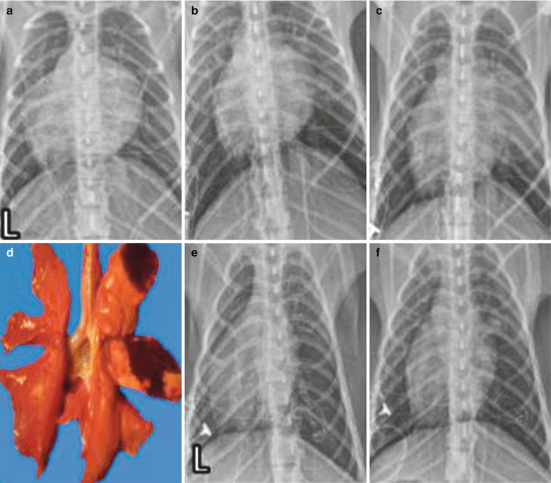
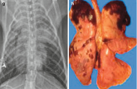


Fig. 19.2
Pneumonic plague. (a) Chest X-ray demonstrates clear pulmonary markings in both lungs in African green monkeys (X775) before their infection of Yersinia pestis; (b) chest X-ray demonstrates pale flakes of shadows in the right middle lung lobe at day 3 after the infection; (c) chest X-ray demonstrates flakes of high-density shadows in the right middle lung lobe and pale shadows in the right upper lung lobe 4 days later; (d) autopsy of the gross specimens after euthanasia demonstrates flakes of necrotic areas in the right middle and upper lung lobes; (e) chest X-ray demonstrates clear pulmonary markings in both lungs in African green monkeys (X784) before their infection of Yersinia pestis; (f) chest X-ray demonstrates pale flakes of shadows in the right middle lung lobe at day 3 after the infection; (g) chest X-ray demonstrates flakes of pale shadows and high-density shadows in the middle lobes of both lungs and in the left lower lung lobe; (h) autopsy of the gross specimens after euthanasia demonstrates spots and flakes of necrotic necroses areas in the upper lobes and lower lobes of both lungs
(Note: L for left. Reproduced with permission from Layton et al. RC, et al. J Med Primatol, 2011b, 40(1): 6.)
Case Study 3
A male patient aged 53 years complained of high fever with chills and a body temperature of 40 °C, cough with frothy bloody sputum, chest pain, obvious dyspnea, and mild headache. Reverse indirect agglutination test of sputum specimen at day 2 after the onset demonstrated Yersinia-specific F1 antigen positive, moist rales in both lungs, phlegm rales in the left lung, lower breath sounds in the right lung, and dullness on percussion.
For case detail and figures, please refer to DaWa WJ, et al. Chinese Journal of Tuberculosis and Respiratory Disease, 2011, 34(6): 404. (In Chinese)
Case Study 4
A female patient aged 40 years complained of high fever, cough with light yellowish foam-like sputum in small quantity and with blood streaks, chest pain, and breathing difficulty. Her SpO2 was 80 %, with coarse breathing sounds in both lungs and moist rales in the left middle lung. Reverse indirect agglutination test of sputum specimen demonstrated Yersinia-specific F1 antigen positive.
For case detail and figures, please refer to DaWa WJ, et al. Chinese Journal of Tuberculosis and Respiratory Disease, 2011, 34(6): 404. (In Chinese)
Case Study 5
A male patient aged 37 years complained of fever with a body temperature of 39.8 °C, chest pain, slight breathing difficulty, and cough with yellowish thick sputum that is difficult to be coughed up with no blood in it. Extensive moist rales and a little phlegm rales can be heard in the left lung. Reverse indirect agglutination test of sputum specimen demonstrated Yersinia-specific F1 antigen positive.
For case detail and figures, please refer to DaWa WJ, et al. Chinese Journal of Tuberculosis and Respiratory Disease, 2011, 34(6): 404. (In Chinese)
Case Study 6
A male patient aged 20 years complained of fever with a body temperature of 39 °C, slight cough, expectoration with blood streaks, and occasional chest pain. A few moist rales can be heard in the right middle lung. And his conditions are relatively slight. Reverse indirect agglutination test of sputum specimen demonstrated Yersinia-specific F1 antigen positive.
For case detail and figures, please refer to DaWa WJ, et al. Chinese Journal of Tuberculosis and Respiratory Disease, 2011, 34(6): 404. (In Chinese)
Case Study 7
A male patient aged 38 years complained of fever and body temperature of 37.5 °C, cough with frothy bloody sputum, chest pain, obvious dyspnea, fatigue, myalgia, and nausea with vomiting. Reverse indirect agglutination test of sputum specimen at day 3 after the onset demonstrated Yersinia-specific F1 antigen positive, phlegm rales in the right lung, and lower breath sounds in the left lung (Fig. 19.3).
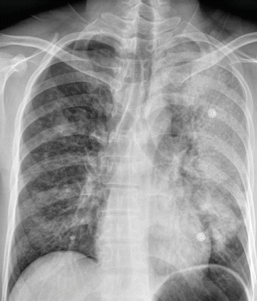

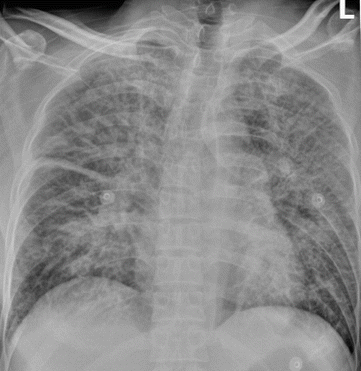
Fig. 19.3
Pneumonic plague. At day 2 of the illness course, chest X-ray demonstrates spotted and flocculent shadows in the right upper lung field and large flakes of shadows in the left lung field
Case Study 8
A male patient aged 50 years with a body temperature of 38.5 °C and obnubilation and had the fidgets and hemorrhage spots in the skin. Bilateral anisocoria, left 2 mm, right 3 mm, light reflex slow. His SpO2 cannot be measured, with coarse breathing sounds in both lungs and some moist rales. Reverse indirect agglutination test of sputum specimen at day 3 after the onset demonstrated Yersinia-specific F1 antigen positive (Fig. 19.4).
