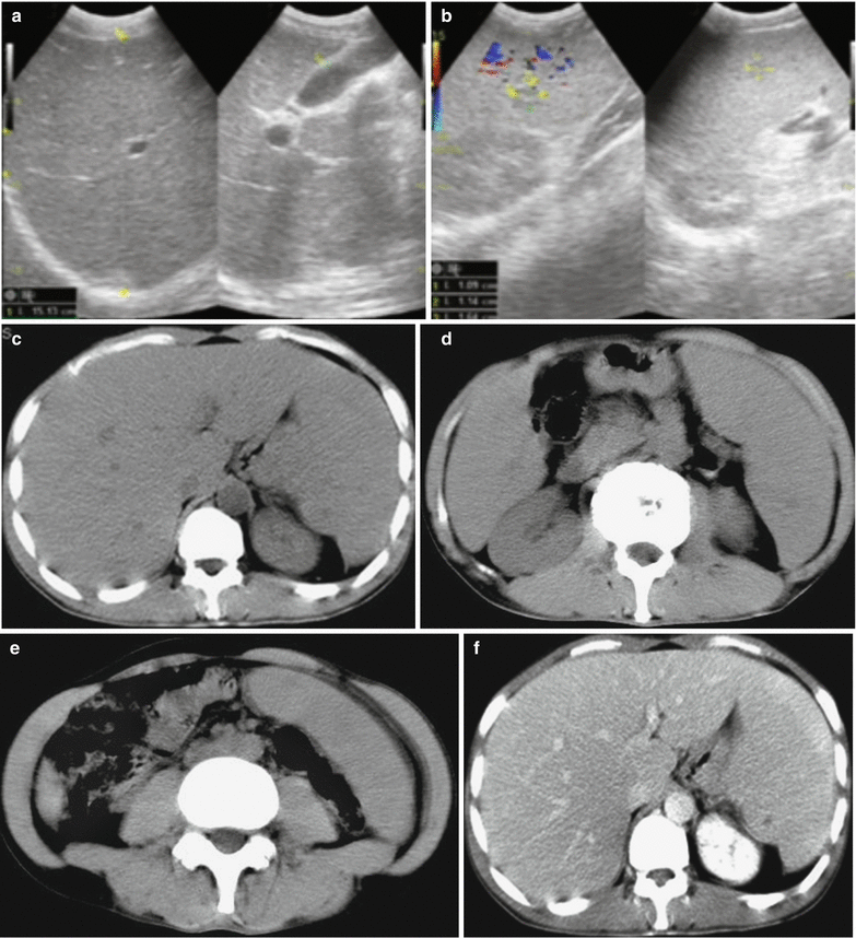
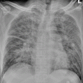
Fig. 27.1
Enlarged spleen in a patient with visceral leishmaniasis. Ultrasound demonstrates enlarged spleen
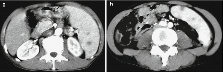
27.7.2 CT Scanning
CT scanning has no specific demonstration, with common findings of enlarged liver and spleen. In some cases, CT scanning can reveal conditions of splenic infarction and liver abscesses (Figs. 27.2, 27.3 and 27.4).
Case Study 2
Visceral leishmaniasis
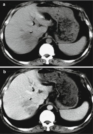
Fig. 27.2
Hepatic lesions of visceral leishmaniasis. (a, b) Plain CT scanning reveals multiple flakes of foci with low density in the right hepatic lobe, with unclearly defined boundaries. Contrast CT scanning demonstrates slightly enhanced foci still in low density
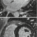
Case Study 3
A male patient aged 43 years was admitted to the hospital due to intermittent fever for 5 months. He had a history of living in an epidemic area of visceral leishmaniasis and a history of mosquito stings and bites. Serological test for visceral leishmaniasis is positive.
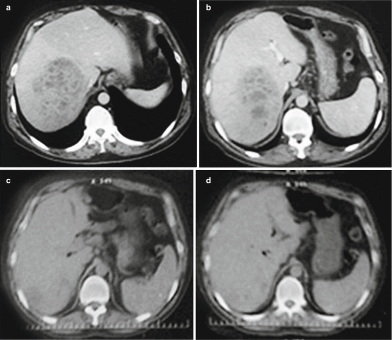
Fig. 27.3
Hepatic lesions of visceral leishmaniasis. (a, b) CT scanning demonstrates large flakes of honeycomb-liked low-density shadow in the right hepatic lobe, with quite clearly defined boundaries and uneven inner density. Contrast CT scanning demonstrates insignificantly enhanced foci. (c, d) CT scanning demonstrates absence of the hepatic lesions in the right hepatic lobe and decreased size of the spleen
Case Study 4
A male patient aged 38 years had persistent fever for 2 months, with a body temperature of 39 °C. He engaged in engineering construction in an area with epidemic of visceral leishmaniasis and had a history of mosquito stings and bites. Serological test for visceral leishmaniasis is positive.