Fig. 15.1
Lyme disease complicated by neurological lesions. (a) T1WI demonstrates strips of low signal in the left thalamus. (b) T2WI FLAIR demonstrates stripes of high signal in the left thalamus
Note: The case and images are provided by Tang, YH. at Rui Jin Hospital, Shanghai, China
15.7.3.2 Joint Lesions
For joint lesion, MR imaging is superior in demonstrating lesions of the soft tissues and joint cartilages. The main findings include bone erosion around the major joints, multiple cystic degenerations under the bone cortex, osteophyte formation, absent joint cartilage, and articular effusion. In some pediatric cases, there are also enlarged lymph nodes in the soft tissues surrounding the joints, myositis, and fasciitis. Contrast T1WI demonstrates enhanced enlarged lymph nodes (>1 cm), flakes of enhancement in the muscles around the joints, and edema around superficial and deep fascia.
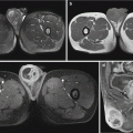
Case Study 4
A male patient aged 16 years.
For case detail and figures, please refer to Ecklund et al. AJR Am J Roentgenol, 2005, 184 (6): 1904.
15.7.3.3 Myocarditis
In the acute stage, delayed contrast MR imaging demonstrates stripes of enhancement in the myocardium (Fig. 15.2).
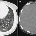
Case Study 5
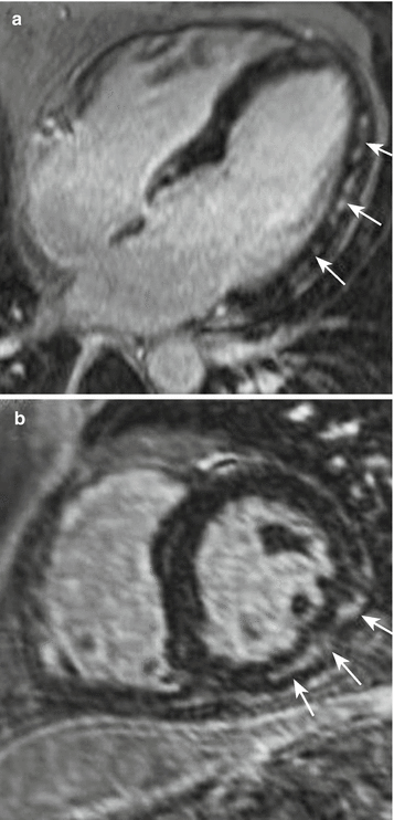
Fig. 15.2
Lyme disease complicated by cardiac lesions. (a) Contrast imaging of the four cardiac chambers in the advanced stage demonstrates moderate enhancement in the left ventricular wall (arrows). (b) Short-axis imaging demonstrates stripes of enhanced lesions in the left ventricular wall (arrows) (Reproduced with permission from Maher B, et al. Heart. 2012, 98 (3): 264)
15.7.4 Nuclear Medicine
Generally, assessment and diagnosis of Lyme disease do not necessarily require nuclear medicine examination. And it fails to demonstrate abnormalities for early neurological lesions. The advanced neurological lesions are usually demonstrated as decreased cerebral cortex perfusion and increased radioactive uptake by the affected joint.
For case detail and figures, please refer to Sumiya H, et al. J Nucl Med, 1997, 38 (7): 1120.
15.8 Diagnostic Basis
The diagnosis of the Lyme disease should be based on the epidemiologic history, clinical manifestations, and laboratory tests.
15.8.1 Epidemiology
The patient has a history of visit or living in an epidemic area in the epidemic season, with an experience of being bitten by a tick.
15.8.2 Clinical Manifestations
The characteristic erythema chronicum migrans and the skin lesions being above 10 cm in diameter highly indicate Lyme disease.
15.8.3 Laboratory Tests
Bb can be isolated from the tissue fluid or body fluid of the patient. One month after the infection, IgG positive supports the diagnosis of Lyme disease. IgG positive alone is likely to be false-positive.
Related posts:

Stay updated, free articles. Join our Telegram channel

Full access? Get Clinical Tree


