(1)
Department of Radiology, Beijing You’an Hospital, Capital Medical University, Beijing, China
Psittacosis, also known as ornithosis, is an acute infectious disease caused by Chlamydia psittaci (Cps) and commonly prevails in poultry and other species of bird. Humans infected by Chlamydia psittaci may suffer from unapparent subclinical infection, with symptoms ranging from mild flulike illness to severe SARS. As a typical animal-based infectious disease, psittacosis rarely has pulmonary signs but a long illness course, despite its clinical manifestation characterized by severe pulmonary lesions. Repeated onsets of psittacosis may lead to chronic diseases.
20.1 Etiology
Initially isolated from parrots, Chlamydia psittaci (Cps) is the pathogen of psittacosis. With a diameter of 150–200 nm, the elementary body is ring-shaped and characterized by a narrow protoplasmic margin around the nucleoplasm, a non-glycogen inclusion body and iodine staining negative. Cps develops well in several cell culture systems, among which HeLa cells, Vero cells, and L cells as well as McCoy cells are commonly used. The Cps can also develop in the yolk sac of the chicken embryo. The number of susceptible animals is relatively large, and the laboratory rats are usually used in the animal inoculation. As Cps and Chlamydia trachomatis share the same antigen, both of them cannot be distinguished by the complement fixation test (CFT). With a weak resistance to the surroundings, Cps can be easily killed by the general chemical disinfectants. It can be inactivated in 48 h at 37 °C, in 10 min at 60 °C, in 24 h with 0.1 % formaldehyde or 0.5 % phenol, and in 30 min with diethyl ether or with ultraviolet radiation. It is resistant to low temperature and can remain infectious for several years if it is kept at –70 °C.
20.2 Epidemiology
20.2.1 Source of Infection
Birds which are infected by psittacosis or serve as the pathogen carriers are considered as the source of infection. Currently, more than 140 types of birds are known to contract or carry the pathogen which is mostly found in secretions and feathers. Infections in birds are unapparent and the signs are characteristic. Although most of the infected birds show no or mild symptoms, the pathogens can be excreted for several months. A patient can also become a minor source of infection if he/she excretes pathogens in sputum.
20.2.2 Route of Transmission
Psittacosis can be transmitted via the respiratory tract. Besides being directly transmitted to humans via droplet, the bacteria can be indirectly transmitted by inhaling an aerosol of infected birds’ feces via the respiratory tract. However, according to the reports, few patients experience the onsets without the contact history of birds. Psittacosis is rarely transmitted via direct person-to-person contact.
20.2.3 Susceptible Population
Populations are generally susceptible and the occurrence has no significant gender difference. It is an epidemic disease all year round. The infection rate is closely related to the frequency of bird contact: parrot and poultry raisers easily contract the disease. Although certain immunity can be acquired after the infection is cured, it is not strong enough to prevent the repeated onsets and the following infection.
20.2.4 Epidemic Features
Psittacosis affects many areas around the world. Although severe outbreaks have occurred, few cases have been reported in recent years. Ritter reported the case of human psittacosis in Switzerland in 1879. In the early 1960s, psittacosis was proved to exist in China. The disease usually spread sporadically, while outbreaks and epidemics occur on a small scale sometimes. As people who are often exposed to poultry and birds have a higher infection rate, psittacosis is considered as an occupational disease for poultry workers.
20.3 Pathogenesis and Pathological Changes
20.3.1 Pathogenesis
The pathogenesis of psittacosis is still not clear. After being inhaled by humans via the respiratory tract, Cps enters the blood circulation, then attacks the mononuclear macrophage system, and proliferates inside. After the above process, Cps invades the lung and other organs via blood. Thus, human psittacosis may be a respiratory tract infection (RTI) or a general infection mainly with respiratory symptoms. The pulmonary lesions often occur in the hilum of the lung and then spread around, resulting in the lobular pneumonia and interstitial pneumonia. Via microscopic examination, infiltration of the mononuclear lymphocytes can be discovered in the alveolar wall and interstitial tissue; deciduous epithelial cells, fibrous protein, lymphocytes, and few neutrophils can be found in the pulmonary alveoli; the inclusion bodies and pathogens can be seen in the cytoplasm of the macrophage in the exudation of pulmonary alveoli. SARS and lobular pneumonia often occur. In severe cases, necrosis of lung tissue may occur accompanying pleurisy. Beside the lung, relative lesions can be found in other involved organs such as the liver, spleen, kidney, meninges, cardiac muscles, and endocardium.
20.4 Clinical Symptoms and Signs
The incubation period generally lasts for 7–40 days, with symptoms ranging from mild to severe. Documents and reports show that the shorter the incubation period, the more severe the symptoms. Mild cases can lead to flulike illness or even no apparent symptoms, while severe cases can result in death. Although SARS is considered as the major manifestation, there is no specific clinical manifestation of psittacosis. Manifestations can be divided into the following types based on the clinical symptoms.
20.4.1 Pneumonia
20.4.1.1 Fever and Flulike Symptoms
The onset is acute, with body temperature up to 40 °C in 1–2 days, chills, general upset, headache, muscular soreness, and obvious muscle shapes in the neck and back. In some cases, patients may suffer from conjunctivitis, epistaxis, and rash. The high body temperature may gradually decline in 1–2 weeks. The fever course is 3–4 weeks, while it may last for several months in some cases.
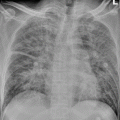
20.4.1.2 Pneumonia
Pneumonia often occurs after the 1st week of onset, accompanying worsening coughs, which are mostly dry coughs with a small amount of phlegm or bloody sputum, choking sensation in chest, and chest pain. In severe cases, patients may suffer from dyspnea, cyanosis, tachycardia, delirium, and even coma. The physical signs of the lung are often milder than the symptoms. At the beginning stage of the onset or in mild cases, patients have no apparent physical signs or only suffer from moist rales in the lung and pharyngeal hyperemia. As the disease develops, signs of lung consolidation and moist rales occur, accompanying pleural friction rub and pleural effusion in few cases.
20.4.1.3 Other Symptoms
Digestive symptoms such as anorexia, nausea and vomiting, abdominal pain, and diarrhea may occur. The liver and spleen may be enlarged accompanying the occurrence of jaundice. The onset of myocarditis, endocarditis, and pericarditis may be triggered, with circulatory failure and pulmonary edema in the severe cases. Psychiatric symptoms such as headache, insomnia, slow response, and nervousness may occur, with sleepiness, delirium, mental disorientation, and unconsciousness in severe cases. The disease can result in serious conditions and adverse outcomes.
The above symptoms are lack of the specificity. The manifestations of pneumonia as well as the enlargement of the spleen play the most important role in the diagnosis of psittacosis.
20.4.2 Typhoid Sepsis and Blood Poisoning
The symptoms include high fever and headache, accompanying relative bradycardia and enlargement of the spleen. Complications such as myocarditis, endocarditis, and meningitis easily occur. In severe cases, the coma and renal failure may lead to patients’ death.
The illness course is long, with a fever course lasting for 3–4 weeks or even several months. The recurrence rate may reach 20 %.
20.5 Psittacosis Related Complications
The complications of psittacosis seldom occur. In severe cases, the cardiovascular system and nervous system can be negatively affected. Cardiac complications include myocarditis, endocarditis, and pericarditis. Cps may attack the aortic valves and mitral valves as well. The disease may lead to artery embolization and have a negative effect on the liver, kidney, skin, and other organs.
20.6 Diagnostic Examinations
20.6.1 Laboratory Tests
20.6.1.1 Routine Tests
At the acute stage, the WBC count is normal or slightly lower, with a normal or slightly accelerating ESR. Transient proteinuria can be discovered via the uronoscopy.
20.6.1.2 Etiological Tests
Etiological tests serve as the diagnostic basis of psittacosis. In clinical cases, blood, sputum, and throat swab are taken at the acute stage to detect Cps.
Related posts:

Stay updated, free articles. Join our Telegram channel

Full access? Get Clinical Tree


