• Y-shaped configuration to lumbar thecal sac on axial imaging
• Mass effect on thecal sac and nerve roots
• Follows fat signal intensity on all sequences
Fat suppression to confirm adipose tissue, exclude blood products
Top Differential Diagnoses
• Subacute epidural hematoma
• Spinal angiolipoma
• Epidural metastasis
• Epidural abscess
Pathology
• Exogenous or endogenous steroids
• General obesity
• Idiopathic
Clinical Issues
• Gradual progression of symptoms
• Weakness: > 85%
• Back pain, sensory loss, polyradiculopathy, altered reflexes, incontinence, ataxia
(Left) Sagittal graphic illustrates confluent abundant epidural fat in dorsal thoracic canal, with effacement of the dorsal thecal sac and mild mass effect on spinal cord, which is displaced ventrally.
(Right) Sagittal T2WI MR of thoracic spine shows smooth and homogeneous dorsal epidural tissue , isointense to subcutaneous fat. Vertebral compression fractures are due to steroid-induced osteoporosis.
(Left) Sagittal T1 MR shows abundant epidural fat both dorsal and ventral to the thecal sac. The thecal sac is small in AP diameter due to compression by the fat.
(Right) Axial T1 MR shows marked prominence of the epidural fat in the lumbar canal, which causes the thecal sac to assume a trefoil pattern. The abundant fat in the epidural space allows for good visualization of the normal anatomy of the Hoffman ligament, which connects the posterior longitudinal ligament to the ventral dura .
TERMINOLOGY
Definitions
• Excessive accumulation of intraspinal fat causing cord compression and neurologic deficits
IMAGING
General Features
• Best diagnostic clue
Abundant epidural fat in midthoracic and distal lumbar spinal canal compressing thecal sac
• Location
Thoracic spine: 58-61%
– T6-T8, dorsal to spinal cord
Lumbar spine: 39-42%
– L4-L5, surrounding thecal sac
• Size
Epidural fat ≥ 7 mm thick in thoracic spine
Over multiple vertebral segments
• Morphology
Y-shaped configuration to lumbar thecal sac on axial imaging


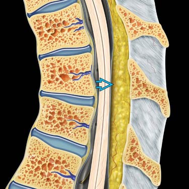
 in dorsal thoracic canal, with effacement of the dorsal thecal sac and mild mass effect on spinal cord, which is displaced ventrally.
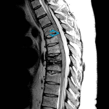
 , isointense to subcutaneous fat. Vertebral compression fractures are due to steroid-induced osteoporosis.
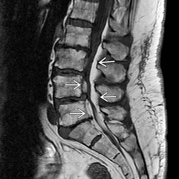
 both dorsal and ventral to the thecal sac. The thecal sac is small in AP diameter due to compression by the fat.
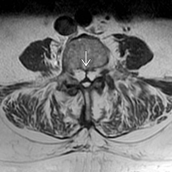
 .
.


