Etiology
Erectile dysfunction manifests clinically most commonly as impotence and less commonly as priapism. The causes of impotence can be psychogenic, endocrinologic, neurogenic, anatomic, infectious, pharmacologic, or vasogenic. Vasogenic causes of erectile dysfunction include venous leak (aging, priapism, congenital, idiopathic) and arterial insufficiency (atherosclerosis, arterial stenosis or occlusion, perineal radiation, iatrogenic). Anatomic causes include phimosis and paraphimosis and fibrous plaques/scarring (from ischemia, Peyronie’s disease, scleroderma, and chronic priapism). Priapism is an uncommon medical condition. Anatomic and vascular causes of erectile dysfunction are curable by surgery and thus their accurate detection and characterization are clinically important.
Prevalence and Epidemiology
With growing societal openness toward sexual dysfunction, more men are coming forward for evaluation and management of erectile dysfunction. Traditionally, male impotence was considered to be psychogenic or an inevitable consequence of aging. However, recent research indicates that arterial, venous, or mixed arteriovenous insufficiency plays a role in most cases of impotence. Frequently, impotence is caused by a combination of psychogenic and organic factors.
Clinical Presentation
Impotent patients report inability to achieve penile erection, in contrast to priapism, in which patients present with persistent penile tumescence that is not associated with sexual desire or stimulation. Other clinical manifestations related to causal or associated disease may predominate in these patients.
Pathophysiology
Penile erection is a neurovascular phenomenon resulting from arterial dilatation, sinusoidal relaxation, and venous outflow restriction. Arterial compliance and adequate blood inflow are essential to achieve erection, and restriction of venous outflow is essential to preserve it. Parasympathetic nerve stimulation via the S2-S4 nerve roots causes erection of the penis, and sympathetic nerve stimulation via the T11-L2 roots causes its detumescence.
During erection, cholinergic stimulation causes relaxation of the smooth muscles surrounding the lacunar spaces of corpora cavernosa. This reduces peripheral vascular resistance and increases arterial inflow. Relaxation of the smooth muscles of the cavernous and helicine arteries causes their dilatation. When the sinusoids are expanded and engorged with blood, emissary veins are mechanically compressed against the unyielding tunica albuginea, which helps to maintain sinusoidal distention and penile erection.
During detumescence, sympathetic neural activity causes contraction of the smooth muscles of the helicine arteries and the lacunae with a resultant decrease in arterial inflow. This results in collapse of the lacunae, with loss of erection.
Pathology
Organic impotence is most commonly due to vascular pathologic processes. Venous incompetence or failure of the veno-occlusive mechanism of outflow restriction has been recognized as a major factor and may be its most common cause. Venous leakage may be spongiosal and may result from deterioration of the tunica albuginea, which is worsened by repeated and prolonged erections as a result of high pressure in the corpora cavernosa. Venous leakage also may result from aging or priapism or be congenital or idiopathic. Both the cavernosal and para-arterial veins play an important role in the circulation of the corpora cavernosa and may be responsible for recurrence of venous leakage after deep dorsal venous stripping surgeries. Arterial disease within the proximal pelvic or segmental penile arteries can result in insufficient arterial inflow. Arterial insufficiency may occur in isolation or be accompanied by venous leakage. In patients older than 50 years of age, arterial insufficiency is usually caused by atherosclerosis affecting the iliac or internal pudendal arteries. In young patients, traumatic focal stenosis or occlusion of the common penile artery in the region where it courses directly over the ischiopubic rami may result in erectile dysfunction. Perineal radiation therapy may affect the common penile artery close to the prostate gland.
Other, less common causes of erectile dysfunction include anatomic abnormalities such as phimosis and paraphimosis. Penile fibrosis within or adjacent to the tunica albuginea may cause painful erection, whereas fibrosis in the corpora cavernosa may prevent lacunar filling. There are multiple causes of penile fibrosis, including ischemia, Peyronie’s disease, and scleroderma. Ischemic injury secondary to chronic priapism may result in fibrotic scarring and, hence, erectile dysfunction. Priapism is most commonly caused by intrapenile injections of vasoactive drugs or by hypercoagulable states, including sickle cell disease and disseminated malignancy. Prolonged high-flow priapism secondary to arteriosinusoidal fistula (resulting from perineal or penile trauma) may also result in cavernosal ischemia, fibrosis, and erectile dysfunction.
Imaging
The penile-brachial index was used in the past for evaluation of vasculogenic impotence. However, this index may be affected by aortoiliac disease, even when penile hemodynamics are normal. Techniques for evaluation of erectile dysfunction include duplex Doppler and color Doppler ultrasonography, dynamic infusion cavernosometry and cavernosography, and arteriography, including selective pudendal pharmacoangiography. At present, penile Doppler ultrasonography is the mainstay of imaging evaluation of impotence.
Radiography
Cavernosometry is usually performed in patients for whom venous surgery is being considered. The corpora cavernosa are cannulated with 25-gauge butterfly needles. Warm saline and low osmolar contrast material is infused via one corpus cavernosum, perfusing the other via anastomotic connections. Direct pressure measurements are obtained via the needle in the other corpus cavernosum. After baseline measurement of intracavernosal pressure and penile circumference, pharmacologic erection is produced by intracavernosal injection. Pressure and penile circumference monitoring is then performed for 10 minutes or until equilibrium occurs. If no erection is produced, heparinized saline is infused at increasingly rapid rates until intracavernosal pressure of 150 mm Hg is achieved. An infusion rate of less than 30 mL/min is considered normal. Venous outflow resistance also may be assessed by observing the rate of intracavernosal pressure fall after termination of intracavernosal saline infusion. With venous leakage, pressure falls rapidly. In cavernosography, 100 to 150 mL of contrast agent is infused into the corpus cavernosum to maintain 90 mm Hg of intracavernosal pressure. Fluoroscopy and spot films during the procedure may reveal the site of venous leak and provide the preprocedural anatomic information.
Fibrous plaques and calcifications resulting from Peyronie’s disease or scleroderma may be seen as filling defects within the corpora on cavernosography.
Selective internal pudendal pharmacoarteriography is usually performed before reconstructive arterial surgery or angioplasty. Intracavernosal injection of papaverine, nitroglycerin, or prostaglandin E1 (PGE 1 ) is administered during the procedure. Aortoiliac arteriography is performed to evaluate for proximal atherosclerotic lesions and the patency of the inferior epigastric arteries, which are usually used for surgical revascularization.
Magnetic Resonance Imaging and Computed Tomography
MRI and CT are useful alternatives for evaluation of arteriogenic impotence secondary to aortoiliac or small vessel disease and can be useful for evaluation of priapism secondary to venous thrombosis. It also can detect fibrotic plaques such as those resulting from Peyronie’s disease and scleroderma. When calcified, these plaques can be identified on CT.
Ultrasonography
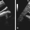
The ultrasound evaluation of erectile dysfunction uses Doppler estimation of cavernosal artery velocities to characterize arterial inflow. A high-frequency linear transducer is used with a mechanical standoff wedge to produce a favorable insonating angle. Optimization of “slow flow” sensitivity is crucial for the accurate depiction of diastolic flow and blood flow in the dorsal vein. Ultrasound evaluation of the flaccid penis is first performed to assess for structural anomalies and plaques. Color Doppler imaging is used to identify the cavernosal artery before and after erection and determine its location and the flow direction. Pharmacologic enhancement of erection can be achieved by injecting 30 to 60 mg of papaverine intracavernosally near the penile base. Other agents used to induce erection include phentolamine and PGE 1 . Intracavernosal injection of PGE 1 is contraindicated in men who have conditions that predispose to priapism, such as sickle cell anemia, sickle cell trait, multiple myeloma, leukemia, cavernosal thrombosis, and tumors that are known to invade the cavernosa. Use of PGE 1 is also contraindicated in men with penile prostheses and relatively contraindicated in men with anatomic abnormalities of the penis. The diameter of the cavernosal artery may be immeasurably small, at 0.6 mm in diameter before erection ( Figure 75-1, A ), and increases more than 75% in diameter, to an average of 1 mm in the erect penis (see Figure 75-1, B ). Both cavernosal arteries are intermittently imaged with Doppler ultrasonography at 5-minute intervals from 1 to 25 minutes after pharmacologic induction of erection or until waveform progression ceases. The normal peak systolic velocity in the erect penis ranges from 35 to 60 cm/s. In the flaccid penis, Doppler ultrasonography demonstrates monophasic flow with minimal diastolic flow ( Figure 75-2, A ). With the onset of erection or after papaverine injection, there is an increase in both systolic and diastolic flow (see Figure 75-2, B ). As the intracavernosal pressure increases, a dicrotic notch appears at the end of systole accompanied with the decrease in diastolic flow. With further increases in intracavernosal pressure, end-diastolic flow diminishes to zero and then reverses ( Figure 75-3 ). Thus, a high-resistance waveform should be seen in the cavernosal artery at erection, with little or no diastolic flow. Peak systolic velocities of 11 to 20 cm/s and over 35 cm/s are typical in the normal cavernosal and dorsal arteries before and after intracavernosal PGE 1 injection. The venous outflow occlusive mechanism is most functional at full penile rigidity with adequate arterial inflow. Consequently, a high-resistance waveform with little or no diastolic flow should be seen in the cavernosal artery at erection.


