Fig. 2.1
Candida esophagitis in a 73-year-old male patient. (a) On double-contrast esophagogram, diffuse nodularity is seen in the esophagus. (b) Multiple whitish plaques are noted on endoscopic examination
2.10.2 Herpes Esophagitis in an Immunocompetent Patient
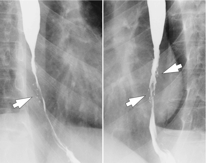
Fig. 2.2
Herpes esophagitis in a 23-year-old immunocompetent male patient. Multiple small punctate ulcerations are seen in the distal esophagus (arrows). This finding is characteristic of herpes esophagitis in immunocompetent patients. Note the diffuse luminal narrowing in the distal esophagus due to underlying eosinophilic esophagitis
2.10.3 Cytomegalovirus Esophagitis
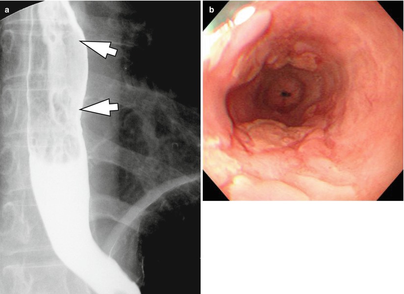
Fig. 2.3
Cytomegalovirus esophagitis in a 57-year-old female patient. (a) Multiple large nodular lesions with central superficial ulceration are seen in the distal esophagus (arrows). (b) Multiple nodularities and superficial ulcerations with elevated margin are also seen on endoscopic examination
2.10.4 Reflux Esophagitis with a Focal Stricture
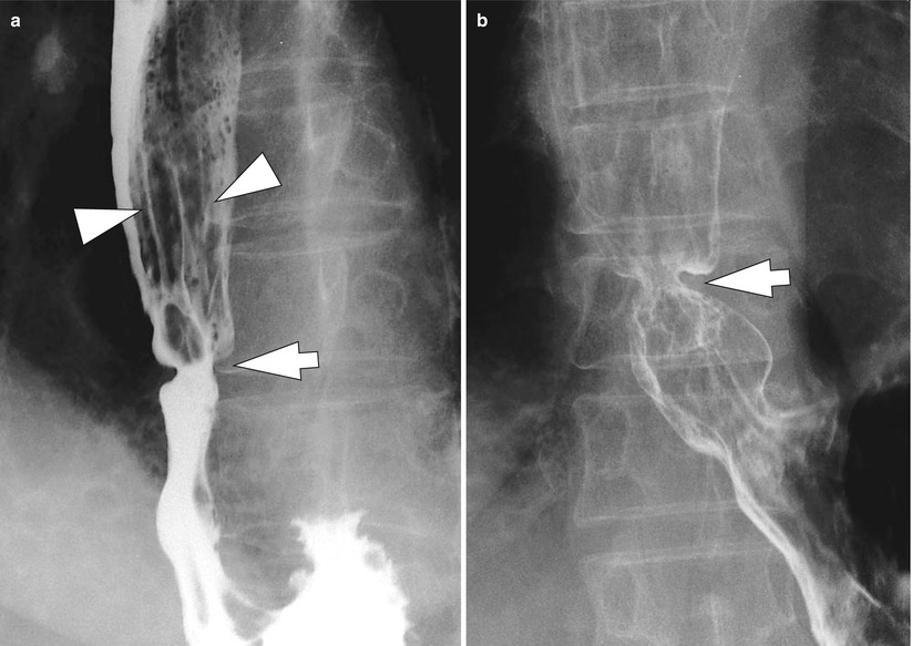
Fig. 2.4
Reflux esophagitis with a focal stricture in a 63-year-old female patient. (a) A focal stricture (arrow) is seen in the distal esophagus with proximal esophageal dilatation and adjacent fold thickening (arrowheads). (b) Double-contrast esophagogram well demonstrates focal esophageal stricture (arrow)
2.10.5 Reflux Esophagitis with Segmental Narrowing
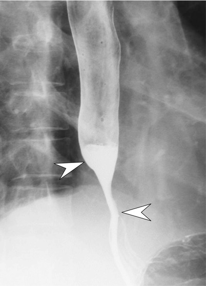
Fig. 2.5
Reflux esophagitis with segmental narrowing in a 50-year-old male patient. Smooth segmental narrowing is noted in the distal esophagus without mucosal lesion such as ulceration (arrowheads). This finding suggests reflux esophagitis more likely than tumorous condition
2.10.6 Drug-Induced Esophagitis Caused by Alendronate Sodium
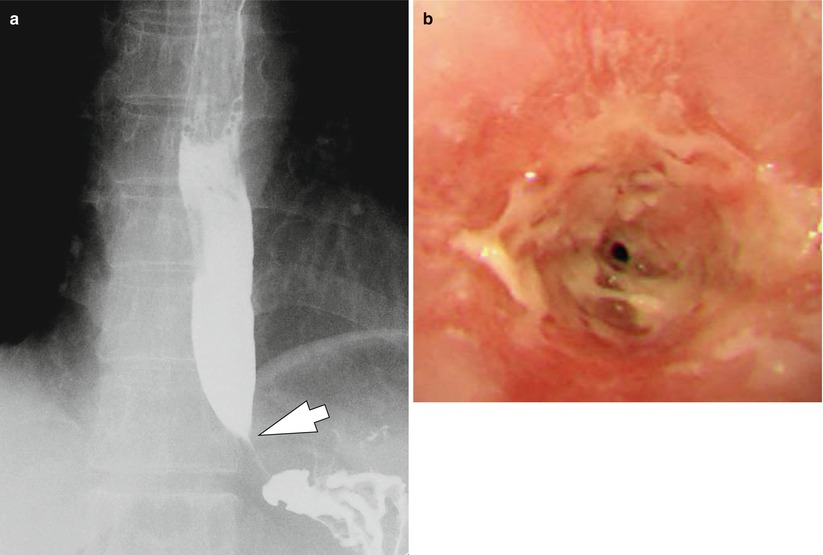
Fig. 2.6
Drug-induced esophagitis caused by alendronate sodium in a 61-year-old female patient. (a) Luminal narrowing (arrow) is seen in the distal esophagus, adjacent to lower esophageal sphincter. (b) There is an esophageal stricture at the level of 38 cm from incisor, so that the endoscopic scope couldn’t pass through it. This patient was taking alendronate sodium (Fosamax) for the treatment of osteoporosis
2.10.7 Acute Radiation Esophagitis
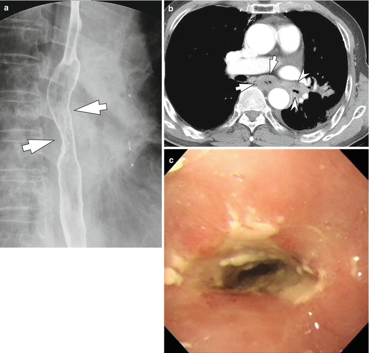
Fig. 2.7


Acute radiation esophagitis in a 69-year-old male patient receiving concurrent chemoradiation therapy for non-small cell lung cancer. The patient complained dysphagia and odynophagia a few weeks after starting concurrent chemoradiation therapy. (a) On double-contrast esophagogram, mucosal nodularity and ulceration are seen in the esophagus at the subcarinal level (arrows). Decreased distensibility of the irradiated segment is also noted. (b) Contrast-enhanced CT image demonstrates segmental esophageal wall thickening at the subcarinal level (arrows). The residual enhancing consolidative mass in the left lower lobe of the lung (arrowheads) is also seen. (c) The endoscopic examination shows luminal narrowing and whitish mucus with ulcerative lesion in the esophagus at the level of 25 cm from incisor
Related posts:

Stay updated, free articles. Join our Telegram channel
Full access? Get Clinical Tree
