Fig. 3.1
The transducer is the most fragile part of the ultrasound unit and should be protected from drop damage during storage and scanning
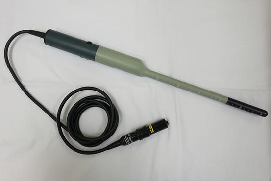
Fig. 3.2
During sterilization of intracavity transducers the transducer coupling should be protected
Setting Up the Ultrasound Room
The physical environment in which pelvic floor ultrasound is performed can have significant impact on a patient’s or sonographer’s comfort and indirectly affect the quality of images and interpretation of the scan findings. This is especially because pelvic floor imaging is often performed as part of functional/dynamic assessment of pelvic floor dysfunction.
Both the examination table and the sonographer’s chair should ideally be height adjustable. The physical configuration of the scanning room should ensure that the clinician has ready access to the ultrasound machine, examination table, gel, towels and lighting controls (Figs. 3.3 and 3.4). Space for a support person (for the patient) or chaperone is also important especially when performing intracavity imaging.
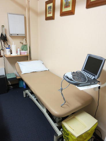
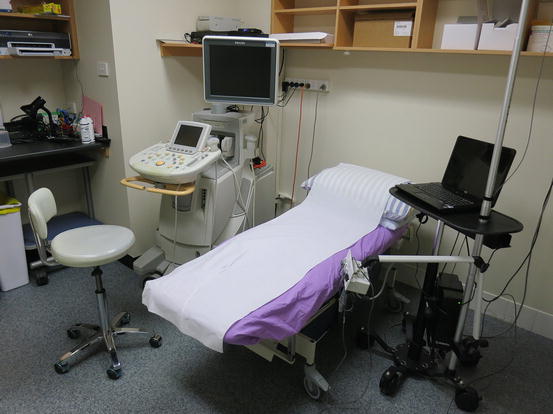
Fig. 3.3
Bedside ultrasound setup in a consultation room
Fig. 3.4
 Practical Application of Ultrasound in the Assessment of Pelvic Organ Prolapse
 Pelvic Ultrasound in the Assessment of Female Voiding Dysfunction
 Machine Settings and Technique of Image Optimization
 Endoanal Ultrasound of Pelvic Floor
 Principles and Applications of 3D Pelvic Floor Ultrasound
 The Physics and Technique of Ultrasound


Dedicated ultrasound room setup for pelvic floor imaging and ultrasound urodynamics
Related posts:
Practical Application of Ultrasound in the Assessment of Pelvic Organ Prolapse
Pelvic Ultrasound in the Assessment of Female Voiding Dysfunction
Machine Settings and Technique of Image Optimization
Endoanal Ultrasound of Pelvic Floor
Principles and Applications of 3D Pelvic Floor Ultrasound
The Physics and Technique of Ultrasound
Stay updated, free articles. Join our Telegram channel
Full access? Get Clinical Tree
