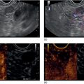
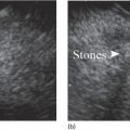
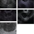
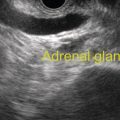
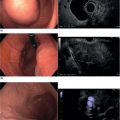
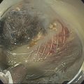
Michael Chang Department of Medicine, University of California San Diego, La Jolla, CA, USA Achalasia is characterized by impaired deglutitive relaxation of the lower esophageal sphincter (LES), considered to be a result of neuronal degeneration in the myenteric plexus and consequently an imbalance between excitatory and inhibitory neurons. Achalasia can present at any age, with a peak incidence between 30 and 60 years, without a racial or gender predilection. The etiology of achalasia is typically idiopathic, but can also be associated with diseases such as Chagas, sarcoidosis, or malignancies. Progressively worsening dysphagia for both liquids and solids is present in over 90% of patients with achalasia. Other symptoms may include chest fullness, heartburn, regurgitation, early satiety, and weight loss. High‐resolution esophageal manometry is the gold standard for diagnosing and classifying achalasia. An elevated median integrated relaxation pressure (IRP) along with absent peristalsis is suggestive of type I achalasia (classic achalasia), or of type II achalasia when accompanied with panesophageal pressurization. In type III achalasia, esophageal contractility will be spastic in the setting of an elevated median IRP. Barium esophagram, a helpful complementary diagnostic test, may demonstrate a dilated esophagus with a classic “bird’s beak” appearance in type I or II achalasia. In the case of type III achalasia, corkscrewing in the esophageal body may be seen on barium esophagram. Functional lumen imaging probe is another complementary diagnostic tool in achalasia, in which distensibility at the esophagogastric junction is reduced and contractile response to distension in the esophageal body is abnormal. All patients with a diagnosis of achalasia or suspected achalasia should undergo upper endoscopy to confirm the diagnosis and rule out esophageal malignancy.
10
EUS for Achalasia
Introduction
Clinical presentation and diagnosis
Role of EUS in achalasia
Stay updated, free articles. Join our Telegram channel

Full access? Get Clinical Tree


