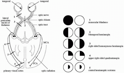
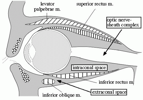
MONOCULAR DEFECTS
1 = monocular blindness (optic nerve lesion in fracture of optic canal, amaurosis fugax)
BILATERAL heteronymous DEFECTS
2 = bitemporal hemianopia (chiasmatic lesion)
BILATERAL HOMONYMOUS DEFECTS
3 = homonymous hemianopia
4 = upper right-sided quadrantanopia
5 = central hemianoptic scotoma
Optic neuritis
Vascular ischemia
Amaurosis fugax = cholesterol emboli from internal carotid artery occluding central retinal artery and its branches
Occult cerebrovascular malformation affecting the optic nerve
Temporal arteritis
Malignant optic glioma of adulthood
INFLAMMATORY DISEASE
Tissue-specific inflammation: orbital cellulitis, optic neuritis, scleritis, myositis, Graves disease
Panophthalmitis
Pseudotumor of orbit
CYSTIC DISEASE
Dermoid cyst
Mucocele
Retroocular cyst (developmental)
VASCULAR LESIONS
arterial and arteriovenous lesion
1. Ophthalmic artery aneurysm
2. Arteriovenous fistula (rare) eg, Wyburn-Mason syndrome
3. Carotid-cavernous fistula
capillary lesion
4. Capillary hemangioma/benign hemangioendotheliloma
venous vascular malformation
5. Cavernous hemangioma
6. Orbital varix
venous lymphatic malformation
7. Capillary lymphangioma
8. Cavernous lymphangioma
9. Cystic lymphangioma
TUMORS
Rhabdomyosarcoma
Optic nerve glioma
Meningioma
Lymphoma
Metastasis
Hemangiopericytoma
Optic nerve glioma
Optic nerve sheath meningioma (10% of orbital neoplasm)
Optic neuritis
Inflammatory pseudotumor (may surround optic nerve)
Sarcoidosis
Intraorbital lymphoma (may surround optic nerve, older patient)
Elevated intracranial pressure
= distension of optic sheath
[check mark] bilateral tortuous enlarged optic nerve-sheath complex
Optic nerve sheath meningioma
Orbital pseudotumor
Perioptic neuritis
Perioptic hemorrhage
Sarcoidosis
Lymphoma/leukemia
Metastasis
Erdheim-Chester disease = systemic xanthogranulomatosis
Cavernous hemangioma
Orbital varix
Carotid-cavernous fistula
Arteriovenous malformation
least common of orbital vascular malformations (congenital, idiopathic, traumatic)
[check mark] irregularly shaped intensely enhancing mass of enlarged vessels
[check mark] associated with dilated superior/inferior ophthalmic vein
Hematoma
Lymphangioma
Neurilemmoma
[check mark] commonly adjacent to superior orbital fissure, inferior to optic nerve
[check mark] local bone erosion
BENIGN TUMOR
Dermoid cyst
Teratoma
<1% of all pediatric orbital tumors
[check mark] ± areas of fat, cartilage, bone
[check mark] expansion of bony orbit ± bone defect
Capillary hemangioma
Lymphangioma
Plexiform neurofibroma
Inflammatory orbital pseudotumor
Histiocytosis X lesion usually arises from bone
MALIGNANT TUMOR
Lymphoma/leukemia
Metastasis
Rhabdomyosarcoma
FROM SINUS
maxillary/sphenoid sinuses are rare locations of origin
Tumor:
squamous cell carcinoma (80%), lymphoma, adenocarcinoma, adenoid cystic carcinoma
Mucocele
Paranasal sinusitis:
♦ Most common cause of orbital infection!
Origin: from ethmoid sinuses (in children), from frontal sinus (in adolescence)
Organism: Staphylococcus, Streptococcus, Pneumococcus
[check mark] preseptal/orbital edema/cellulitis
[check mark] subperiosteal/orbital abscess
[check mark] mucormycosis (in diabetics) destroys bone + extends into cavernous sinus
Cx:
epidural abscess
subdural empyema
cavernous sinus thrombosis
meningitis
cerebritis
brain abscess
FROM SKIN
Orbital cellulitis
FROM LACRIMAL GLAND
[check mark] mass arising from superolateral aspect of orbit
1. | Dermoid cyst | 46% |
2. | Inflammatory lesion | 16% |
3. | Dermolipoma | 7% |
4. | Capillary hemangioma | 4% |
5. | Rhabdomyosarcoma | 4% |
6. | Leukemia/lymphoma | 2% |
7. | Optic nerve glioma | 2% |
8. | Lymphangioma | 2% |
9. | Cavernous hemangioma | 1% |
mnemonic: LO VISHON | ||
Leukemia, Lymphoma | ||
Optic nerve glioma | ||
Vascular malformation: hemangioma, lymphangioma | ||
Inflammation | ||
Sarcoma: ie, rhabdomyosarcoma | ||
Histiocytosis | ||
Orbital pseudotumor, Osteoma | ||
Neuroblastoma | ||
1. | Retinoblastoma | 86.0% |
2. | Rhabdomyosarcoma | 8.1% |
3. | Uveal melanoma | 2.3% |
4. | Sarcoma | 1.7% |
1. | Leukemia | 36.7% |
2. | Sarcoma | 14.3% |
3. | Hodgkin lymphoma | 11.0% |
4. | Neuroblastoma | 9.2% |
5. | Wilms tumor | 6.7% |
6. | Non-Hodgkin lymphoma | 5.6% |
7. | Histiocytosis | 3.9% |
8. | Medulloblastoma | 3.5% |
Abscess
Intraorbital hematoma
Dermoid cyst
Lacrimal cyst
Lymphangioma
Hydatid cyst
Orbital varix
Arteriovenous malformation
Carotid-cavernous fistula
Hemangioma: capillary/cavernous
Blood cyst
Arterial malformation
Glomus tumor
Hemangiopericytoma
Lacrimal gland tumor
Dermoid cyst
Metastasis (breast, prostate, lung)
Lymphoma
Leukemic infiltration of lacrimal gland
Sarcoidosis
Wegener granulomatosis
Pseudotumor
Frontal sinus mucocele
ENDOCRINE
Graves disease (50%)
Acromegaly
INFLAMMATION
Myositis
• rapid onset of proptosis, erythema of lids, conjunctival injection
Location: single muscle (in adults); multiple muscles (in children)
[check mark] enlarged extraocular muscle
[check mark] positive response to steroids
Orbital cellulitis
Sjögren disease, Wegener granulomatosis, lethal midline granuloma, SLE
Sarcoidosis
Foreign-body reaction
TUMOR
Pseudotumor
Rhabdomyosarcoma
Metastasis, lymphoma, leukemia
VASCULAR
Spontaneous/traumatic hematoma
Arteriovenous malformation
Carotid-cavernous sinus fistula
CONGENITAL
Persistent hyperplastic primary vitreous
Coats disease
Coloboma
Congenital cataract
vitreoretinal
Vitreous hemorrhage
Retinal detachment
Choroidal detachment
Endophthalmitis
Retinoschisis
Retrolental fibroplasia
TUMOR
Retinoblastoma
Choroidal hemangioma
Retinal angiomatosis
Melanocytoma
Choroidal osteoma
TRAUMA
BILATERAL with cataract
Congenital rubella
Persistent hyperplastic vitreous
Retinopathy of prematurity
Retinal folds
Lowe syndrome
[check mark] small globe + small orbit
UNILATERAL
Trauma/surgery/radiation therapy
Inflammation with disorganization of eye (phthisis bulbi)
[check mark] shrunken calcified globe + normal orbit
WITHOUT INTRAOCULAR MASS
generalized enlargement
Axial myopia (most common cause)
[check mark] enlargement of globe in AP direction
[check mark] ± thinning of sclera
Buphthalmos
Juvenile glaucoma
Connective tissue disorder:
Marfan syndrome, Ehlers-Danlos syndrome,
Weill-Marchesani syndrome (congenital
mesodermal dysmorphodystrophy),
homocystinuria
[check mark] “wavy” contour of sclera
focal enlargement
Staphyloma
Apparent enlargement due to contralateral microphthalmia
WITH INTRAOCULAR MASS (rare cause for enlargement)
with calcifications:
Retinoblastoma
without calcifications:
Melanoma
Metastasis
Open-globe injury
Posttraumatic orbital hematoma
Coloboma
Staphyloma
Trochlear calcifications
= aging-related normal variant/young diabetic patient Location: superomedial orbit
Retinoblastoma (>50% of all cases)
Astrocytic hamartoma
Choroidal osteoma
Optic drusen
[check mark] punctate calcifications near optic disc
Scleral calcifications
in systemic hypercalcemic states (HPT, hypervitaminosis D, sarcoidosis, secondary to chronic renal disease)
Retrolental fibroplasia
Phthisis bulbi
Cause: trauma or infection/inflammation
[check mark] small contracted/shrunken calcified/ossified disorganized nonfunctioning globe
Lens implant
Scleral buckle
[check mark] radiopaque/radiolucent device at midglobe level
Intraocular silicone oil injection
[check mark] silicone-related chemical shift artifact
Pneumatic retinopexy
[check mark] gas within globe
Globe prosthesis
Uveal melanoma
Metastasis
86% of ocular lesions within globe; usually in vascular choroid
[check mark] bilateral in 30%
Choroidal hemangioma
Vitreous lymphoma
[check mark] diffuse ill-defined soft-tissue density
Developmental anomalies
Primary glaucoma = enlargement of eye secondary to narrowing of Schlemm canal
Coloboma
Staphyloma
numerous irregular, poorly defined low-intensity echoes:
echogenic material moving freely within vitreous chamber during eye movement
voluminous hyperechoic fibrin clots not fixed to optic nerve (DDx to retinal detachment)
Retinoblastoma
Persistent hyperplastic primary vitreous
Coats disease
Norrie disease
Retrolental fibroplasia
Sclerosing endophthalmitis
TUMOR
Retinoblastoma (most common cause — 58%)
Retinal astrocytic hamartoma (3%):
associated with tuberous sclerosis + von Recklinghausen disease
Medulloepithelioma (rare)
DEVELOPMENTAL
Persistent hyperplastic primary vitreous (2nd most common cause in 19-28%)
Coats disease (4-16%)
Retrolental fibroplasia (3-5%)
Coloboma of choroid/optic disc (11%)
INFECTION
Uveitis
Larval endophthalmitis/granulomatosis (7-16%)
DEGENERATIVE
Posterior cataract (13%)
TRAUMA
Retinopathy of prematurity (5-13%)
Organized vitreous hemorrhage
Long-standing retinal detachment
CALCIFIED MASS
Retinoblastoma
Retinal astrocytoma
NONCALCIFIED MASS
Toxocara endophthalmitis
Coats disease
TUMOR:
Optic nerve glioma
Optic nerve sheath meningioma
Infiltration by leukemia/lymphoma
FLUID:
Perineural hematoma
Papilledema of intracranial hypertension
Patulous subarachnoid space
INFLAMMATION:
Optic neuritis
Sarcoidosis
fusiform thickening
= lens-shaped thickening of nerve-sheath complex
with central lucency: meningioma
without central lucency: optic nerve glioma
excrescentic thickening
= single/multiple nodules along nerve-sheath complex usually due to tumor
tubular enlargement
= uniform enlargement of nerve-sheath complex
with central lucency: subarachnoid process (metastases, perineuritis, meningioma, perineural hemorrhage)
without central lucency: papilledema, leukemia, lymphoma, sarcoid, optic nerve glioma
INFLAMMATION
Dacryoadenitis
Mikulicz syndrome
Associated with: sarcoidosis, lymphoma, leukemia
Sjögren syndrome
• decreased lacrimation, xerostomia
Often associated with: rheumatoid arthritis, systemic lupus erythematosus, scleroderma, polymyositis
Sarcoidosis
TUMOR
6 extraocular muscles | ||
medial rectus muscle | ||
inferior rectus muscle | ||
superior rectus muscle | ||
lateral rectus muscle | ||
superior oblique muscle | ||
inferior oblique muscle | ||
Superior ophthalmic vein | ||
axial CT | ||
coronal CT | ||
Optic nerve sheath | ||
retrobulbar | ||
waist | ||
Globe position | ||
behind interzygomatic line | ||
medial: sphenoid body
above: lesser wing of sphenoid = optic strut
below: greater wing of sphenoid
lateral: small segment of frontal bone
nerves: III oculomotor n.
IV trochlear n.
V1 ophthalmic branch of trigeminal n.:
lacrimal nerve
frontal nerve
VI abducens n.
sympathetic filaments of internal carotid plexus
veins: superior + inferior ophthalmic vein
arteries: 1. meningeal branch of lacrimal artery
2. orbital branch of middle meningeal artery
veins: connection between inferior orbital v. + pterygoid plexus
nerve: optic nerve (I)
vessel: ophthalmic a.
fibrous outermost layer
1. Sclera
2. Cornea
pigmented vascular middle layer = uvea (Lat., grape)
3. Ciliary body (anteriorly)
4. Choroid (posteriorly)
Attachment: tethered to sclera by arteries + veins
innermost sensory layer
5. Retina
Attachment: firm at anterior margin (= ora serrata) and posteriorly at optic disk
anterior segment containing
1. Aqueous humor subdivided by iris into:
anterior chamber
posterior chamber
posterior segment containing
2. Vitreous humor
Optic nerve
Ophthalmic artery
Small veins
Congenital/infantile glaucoma
Neurofibromatosis type 1: obstruction of canal of Schlemm by membranes/masses composed of aberrant mesodermal tissue
Sturge-Weber-Dimitri syndrome
Lowe (cerebrohepatorenal) syndrome
Ocular mesodermal dysplasia (eg, Axenfeld or Rieger anomalies)
Homocystinuria
Aniridia
Acquired glaucoma (rare)
Trauma: laceration of ICA within cavernous sinus
penetrating trauma
surgery
Spontaneous: rupture of an intracavernous ICA aneurysm (in atherosclerosis, Ehlers-Danlos syndrome, osteogenesis imperfecta, pseudoxanthoma elasticum)
Dural sinus thrombosis
Atherosclerosis
superior ophthalmic vein (common)
contralateral cavernous sinus
petrosal sinus
cortical veins (rare)
classic triad:
pulsatile exophthalmos
conjunctival chemosis/edema
persistent auscultatory orbital bruit
restricted extraocular movement
decrease in vision due to increase in intraocular pressure (50%)/cranial nerve deficits = indication for emergent treatment
enlarged edematous extraocular muscles
dilatation of superior ophthalmic vein, cavernous sinus, facial veins, internal jugular vein
focal/diffuse enlargement of cavernous sinus
occasionally sellar erosion/enlargement
enlargement of superior orbital fissure (in chronic phase)
stretching of optic nerve
proptosis
arterial flow in cavernous sinus + superior ophthalmic vein
flow voids in cavernous sinus
early opacification of veins of cavernous sinus
retrograde flow through dilated superior ophthalmic v.
lenticular mass of 7.5 (range, 3-11) mm
intense enhancement similar to choroid
focal thickening of posterior wall of globe
ill-defined mass with intense enhancement
hyperintense to vitreus on T1WI (rule)
isointense to vitreus on T2WI (rule)
hyperechoic homogeneous mass
♦ Most common primary intraocular tumor in adults
♦ Most common uveal melanoma (choroid ” class=LK href=”javascript:void(0)” target=right xpath=”/CT{06b9ee1beed5941992c6d97a44cbf92d6b4115f238e1826376bacbe7dc1f6542b2ee8833965d32e7cafd91a39ee5a210}/ID(AB9-M1)”>> iris ” class=LK href=”javascript:void(0)” target=right xpath=”/CT{06b9ee1beed5941992c6d97a44cbf92d6b4115f238e1826376bacbe7dc1f6542b2ee8833965d32e7cafd91a39ee5a210}/ID(AB9-M1)”>> ciliary body)
initially flat growth profile, later becoming elevated, erupting through Bruch membrane to a characteristic mushroom shape
moderate to strong contrast enhancement
strabismus
may present with leukokoria (if retina massively detached = 16% of leukokoria cases)
strabismus, secondary glaucoma, loss of vision
cholesterol crystals at funduscopy
normal-sized/slightly smaller globe
NO focal mass/calcification (HALLMARK)
clumpy particulate echoes in subretinal space (due to cholesterol crystals suspended in fluid)
vitreous + subretinal hemorrhage (frequent)
unilateral dense vitreus (due to proteinaceous subretinal exudate)
linear V-shaped enhancement at anterior margin of subretinal exudate (due to thickened retina composed of telangiectatic + aneurysmal vessels
mild to moderate linear enhancement of retina at ora serrata + of detached retinal leaves
cystic outpouching (= herniation) of vitreus at site of optic nerve attachment
small globe
Stay updated, free articles. Join our Telegram channel

Full access? Get Clinical Tree


