, Jean-Philippe Bault2, Bernard Benoit3 and Gérard Couly4
(1)
Center of women and fetal imaging, Créteil, France
(2)
Center of fetal imaging Ambroise Paré, Les Mureaux, France
(3)
Princess Grace Hospital, Monaco, France
(4)
Department of maxillo-facial surgery, Necker Hospital, Paris, France
Facial tumors can affect various aspects of the face that are most often linked to their anatomopathological origin.
In this chapter, we do not attempt to be exhaustive, but our objective is to describe a few examples of the tumors seen most frequently in prenatal examinations.
5.1 Teratomas
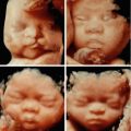
These tumors, also called “epignathus”, have an incidence estimated at between 1/35,000 and 1/200,000 live births. The majority of these tumors originate at the base of the fetal skull and reach the hard palate and the sphenoid. They develop rapidly towards the exterior of the mouth and the nasal cavities, and their extension can be major and impressive, sometimes masking the entire fetal face (Figs. 5.1, 5.2, and 5.3). Sometimes they are more limited (Fig. 5.4).
The appearance of these tumors is most often mixed and disorganized, associating solid and liquid components in variable quantity, without systematic vascularization.
Sometimes these teratomas have a cervical starting point (Fig. 5.5).


Fig. 5.1
Giant epignathus (arrows: ocular structures)
Fig. 5.2
Giant epignathus: HD live rendering highlighting the heterogeneous aspect of the tumor
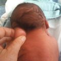
Fig. 5.3
Giant epignathus: post-natal appearance
Fig. 5.4
Small facial teratoma outside of one nostril
Fig. 5.5
Cervical teratoma: heterogenous aspect, vascularization
5.2 Hemangiomas
Vascular anomalies of the face can be divided into vascular malformations and vascular tumors. The most frequent kind of vascular tumor is the hemangioma. These are further divided into those that spontaneously disappear (regression is usually complete by the age of 14 months) and those for which involution is not spontaneous.
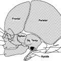
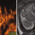
The aspect is most often echogenic, usually homogenous, although sometimes heterogeneous (Fig. 5.7). The Doppler highlights hypervascularization at the level of the tumor (Figs. 5.6 and 5.7). The opposite bone base could be the seat of a slight depression related to the subperiostal process origin (Fig. 5.8).

Fig. 5.6
Frontal hemangioma: left, rendered aspect, right vascularization and slight bone depression