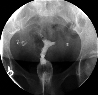
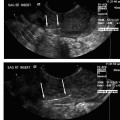
12 Fallopian Tube Recanalization David M. Hovsepian and Gary P. Siskin For nearly two decades, scientific studies have clearly demonstrated the role of selective salpingography and fallopian tube recanalization (FTR) in the evaluation and management of infertile patients.1–23 The American Society for Reproductive Medicine (ASRM) recommends that selective salpingography should be the next step when a diagnostic hysterosalpingogram (HSG) reveals blockage of one or both fallopian tubes.24 However, these techniques remain vastly underutilized for a variety of reasons. The number of FTRs performed annually in the United States represents only a small fraction of the 300,000 or so women who have blockage of their fallopian tubes that should be amenable to catheter-based intervention.25,26 The reasons for this are multiple. Foremost is a lack of familiarity with the ASRM guidelines by referring physicians and radiologists alike. There are also several practical issues related to circumstances and physician preferences. For instance, gynecologists who see infertile patients often perform diagnostic sonography or sonohysterography in their offices and perhaps proceed to in vitro techniques directly, avoiding radiologic tests altogether. Others may recommend laparoscopy from the start, with the goal of investigating any and all possible causes of infertility, buoyed by a handful of articles from a confusing literature. Patients usually have little choice in the matter. Additionally, on the radiology side, FTR may not be available or appropriate in some outpatient settings. Enthusiastic radiologists may find themselves frustrated, even when offering “one-stop shopping,” by insurance, referral patterns, or other issues. In university teaching hospitals, physical and logistical constraints often impede the ability to provide a comprehensive service. A division between diagnostic and interventional services frequently necessitates that FTRs be performed at a separate time and place by radiologists who routinely perform catheter-based procedures. Most interventional radiologists are at least familiar with the techniques involved and many have amassed considerable experience. All should be fundamentally aware, however, of the fertility benefits and low risk of complications. In this chapter, fallopian tube recanalization will be reviewed, from patient selection to techniques, results and outcomes, and potential complications. Our intention is that most readers will readily come to appreciate that the techniques involved are straightforward and can easily be incorporated into practice. The results for this otherwise healthy and well-motivated group of patients can be quite gratifying. Fallopian tube recanalization is not a new or experimental technique. Radiologically speaking, it is prehistoric. In 1849, a London surgeon named W. Tyler Smith was among the first to describe the use of a whalebone bougie to reopen women’s fallopian tubes using only tactile feedback to guide the procedure.27 Over a century would pass before nonsurgical methods to reopen fallopian tubes that had become occluded would again be presented. In 1977, a French radiologist, Dr. J-P Rouanet, published a case report describing the use of standard angiographic equipment to catheterize blocked fallopian tubes.14 This was followed in 1985 by Platia and Krudy, who used a 3F catheter to clear proximally occluded fallopian tubes to treat infertile women.28 A short time later, Dr. Amy Thurmond and her colleagues at the University of Oregon published a series of articles that established the methods and scientific validity of the procedure we know today as fallopian tube recanalization.19,20,29–32 In the years that followed, the list of published series grew and FTR passed beyond the experimental stage into maturity, although many insurance companies still fail to reimburse treatments for infertility, FTR included, based on the perception that tubal occlusion is not truly a disease. Much of the success of FTR rests on the fact that a proximal tubal occlusion is commonly caused by the accumulation of mucus and/or inflammatory debris, although the reason that mucus “plugs” develop remains a source of speculation.33,34 Retrograde menstrual flow can occur35 (which has also been suggested to be a cause of endometriosis, Chlamydia infection,36 and impaired ciliary function37,38 have all been implicated. More recently, work has focused on the influence of hormonally regulated mechanisms that control muscular contractions at the uterotubal junction and ciliary activity. Estrogen and progesterone affect direction and flow of tubal secretions, which may accumulate, inspissate, and eventually calcify.33,34,39 Early intervention may restore a normal luminal interior, whereas chronic blockage can lead to irreversible damage. Once the blockage is cleared, many fallopian tubes appear entirely normal and will resume function and allow conception.40 However, endometriosis, prior surgery and severe pelvic infection can all produce transmural fibrosis and alter normal biological function (fallopian tubes are not just plumbing), so that even after successful recanalization, fertility is still impaired. Primary infertility is defined as the inability to conceive after 12 months of unprotected intercourse. Secondary infertility is the inability to conceive after previously being able to do so, and referred for FTR fall equally into both categories. Patients with secondary infertility often have had prior surgery or instrumentation and generally present more of a technical challenge to recanalization, but by no means should be excluded on this basis. Most women referred for FTR have had a prior HSG documenting occlusion of one or both tubes. Sometimes laparoscopy with chromopertubation (dye injection into the uterus) has demonstrated the blockage. In a study sponsored by the World Health Organization that compared methods for evaluating tubal patency, the findings at laparoscopy and HSG were frequently discordant.41 Nine percent of bilateral occlusions at laparoscopy were found to be patent at HSG, whereas 18% of unilaterally blocked tubes were found to actually be patent. However, false-negatives occurred as well; that is, tubes not visualized at HSG demonstrated spillage of dye at laparoscopy. Overall, the conclusions were that weight of the evidence supported dye injection during laparoscopy as the gold standard to document not only tubal patency, but also for uncovering other significant disease.41 However, HSG remains a useful, low-cost screening tool. Another study, published in 2006, found that the cumulative pregnancy rate did not change whether HSG was performed prior to laparoscopy and intervention, or if patients skipped the HSG and underwent the latter directly.42 Unfortunately, the ASRM guidelines were not followed and no patient underwent selective catheterization after discovery of tubal blockage at the HSG. Therefore, the benefit of transcatheter therapy versus laparoscopic intervention remains an open question. In approximately one-quarter of the patients in that study, endometriosis was found and treated, which is a high proportion relative to the general infertile population, and FTR might be expected to have had less overall success in this group. Over 40% of patients in both arms of the study also underwent adjunctive fertility treatment (intrauterine insemination [IUI) or in vitro fertilization [IVF]), making it difficult to ascertain which aspects of treatment were responsible for the success. Fig. 12.1 This is a single image from a hysterosalpingogram demonstrating a patent right fallopian tube and a proximally occluded left fallopian tube. Although one patent tube alone should be sufficient for conception, the side of ovulation varies month-to-month and the open tube will not regularly correspond to the side bearing the ovum. There are no consistent recommendations regarding what to do when an HSG shows that one tube is patent and the other is not (Fig. 12.1). However, unilateral spill on an HSG can be the result of asymmetric resistance to flow and be an artifact rather than represent an actual obstruction, so one should be careful not to jump to conclusions. Hayashi et al5 evaluated the strategy of proceeding with FTR in 11 patients who had persistent occlusion of only one tube on two successive HSGs. They found that recanalization of the one blocked tube still added benefit, resulting in six pregnancies (55%) on the treated side, confirmed by preovulatory ultrasound examinations identifying a dominant follicle. Hovsepian et al6 also found that having two patent tubes appeared to double the rate of conception, although their study was not sufficiently powered to observe statistical significance nor was the side of conception noted. The patient is placed in the lithotomy position and the pelvis is elevated with a foam cushion or similar padding to allow room to maneuver the speculum (because most fluoroscopy tables cannot accommodate stirrups). The cervix is often located up along the anterior wall of the vagina, not straight ahead, with a posterior orientation that requires the speculum to be angled upward. Elevating the pelvis allows the handle of the speculum to clear the table. The perineum is cleaned with Betadine (Purdue Pharma, Stamford, CT), the patient is draped, and the speculum is inserted. Before cannulation, the cervix is also cleaned with Betadine. The procedure is usually done under sterile technique, with varying adherence to strict guidelines depending on the institution. There are three principal styles of cervical catheterization that employ different equipment, two of which place an acorn-tip catheter on the exterior cervix. One, the Thurmond-Rösch Hysterocath (Cook, Inc., Bloomington, IN), uses a vacuum cup to maintain traction on the cervix, and the other, the Tenacath (Cook, Inc.), secures the cervix with a tenaculum. The third alternative, and there are a variety of products, is a form of balloon-tipped catheter that is advanced across the cervix into the endocervical canal or lower uterine segment. All three styles have their advantages and disadvantages. The vacuum cup is often a challenge to advance through the speculum without contacting the vaginal walls (which can be very uncomfortable) and it is impossible to visualize the cervix as you are advancing it. The axis of the cervix can also deform the cup and interfere with the ability to maintain an adequate vacuum, whether using the manufacturer’s pump or even wall suction. The Tenacath and balloon-tipped catheters have a lower profile and are easier than the vacuum cup to advance through a speculum. Insertion of any of these devices can be greatly facilitated by advancing them coaxially over a catheter and guide wire. The catheter and wire together will help support passage of a balloon-tipped catheter through the cervix or help center an acorn-tipped catheter on the external os. A floppy guide wire, such as a 0.035” Bentson (Cook, Inc.) and an angled 5F catheter, such as a Kumpe or MPA (Cook, Inc.) make a good combination that will pass through any of the above devices. The same combination can also be used for fallopian tube catheterization once inside the uterine cavity. The equipment is preloaded inside the outer cannula before insertion into the cervix. The external os is first engaged with the 5F catheter, then the guide wire is passed up, and the 5F catheter is advanced over the guide wire into the uterine cavity. The catheter and wire serve as the guide for the larger outer cannula. Insertion of a balloon-tipped catheter can be difficult into a nulliparous cervix, and the result can be to push the cervix away and potentially lose access. Often one is only able to position it just inside the endocervical canal. If the balloon is successfully advanced up to the lower uterine segment, gentle traction can be applied that can help to straighten out severe angles. Air is used to inflate the balloon, because saline or contrast will exert greater pressure on the tissues, which can be uncomfortable for hours afterward. Inflation of the balloon is also desirable for sealing the uterine cavity to allow continued visualization during catheterization of the tubal ostia. Occasionally, the angles created by anteversion, retroversion, or flexion will preclude easy cannulation of the tubal ostia. Procedure time may become inadvisably long, translating to increased x-ray dose, so if traction with a balloon-tipped catheter is not an option, the use of a tenaculum is advisable. Using a tenaculum can cause more discomfort for the operator than it does for the patient, especially when anesthetic spray has been applied liberally to the cervix beforehand. Traction on the tenaculum not only gives better visualization of the uterine cavity, it usually makes selective catheterization of the fallopian tubes far easier. The natural shape of the 5F catheter engages the fallopian tube ostia easily and with less discomfort because the uterine fundus is not being stretched by a catheter pressing on it as it loops around. Another advantage of the Hysterocath and Tenacath is the ability to perform a “push—pull” maneuver. By pushing forward and pulling back on the cervix, contrast that has emerged from the tubes into the peritoneal cavity can move to cover the outer surface of the uterus. This is extremely helpful for trying to differentiate a bicornuate from a septate uterus, by demonstrating contrast flowing over a rounded fundal surface or into a cleft between the uterine horns. Once the cervix and endometrial cavity have been accessed, a hysterosalpingogram is performed to delineate the anatomy and identify the cornual regions of the uterus (Fig. 12.2). Not uncommon, proximal fallopian tube occlusion at a prior HSG is no longer evident. This is probably due, in part, to sedatives and narcotics, but can also be attributed to tubal spasm at the time of the initial HSG, which is a well-recognized phenomenon that can account for tubal patency when the examination is repeated at another time. Al-Jaroudi et al1
Background Information
Patient Selection
Technique
![]()
Stay updated, free articles. Join our Telegram channel


Full access? Get Clinical Tree


