Presentation and Presenting Images
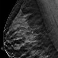
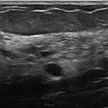
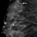
( ▶ Fig. 3.1, ▶ Fig. 3.2, ▶ Fig. 3.3, ▶ Fig. 3.4)
A 40-year-old female presents for baseline asymptomatic screening mammography.
3.2 Key Images
( ▶ Fig. 3.5, ▶ Fig. 3.6, ▶ Fig. 3.7, ▶ Fig. 3.8)
3.2.1 Breast Tissue Density
The breasts are heterogeneously dense, which may obscure small masses.
3.2.2 Imaging Findings
In the upper outer quadrant of the right breast at the 11 o’clock location in the middle depth, there is a circumscribed fat-containing oval mass ( ▶ Fig. 3.5 and ▶ Fig. 3.6). The margins are more conspicuous on the tomosynthesis images ( ▶ Fig. 3.7 and ▶ Fig. 3.8). The left breast is normal ( ▶ Fig. 3.2 and ▶ Fig. 3.4).
3.3 BI-RADS Classification and Action
Category 2: Benign
3.4 Differential Diagnosis
Lipoma: This mass is radiolucent, and a clear thin capsule is seen. A lipoma is a benign neoplasm and is best seen on the digital breast tomosynthesis (DBT) images.
Oil cyst (lipid cyst): An oil cyst is a form of fat necrosis, typically the result of trauma or surgery, and is seen as a radiolucent mass surrounded by a thin capsule that may calcify. DBT may make these more perceptible due to decreased summation artifacts. Summation artifacts occur when fibroglandular tissue components are photographically superimposed to resemble a cancerous lesion. The nature of viewing slices of the breast tissue with DBT reveals that this photographic artifact is composed of layers of normal structures and not a real lesion.
Hamartoma: These benign tumors are composed of various amounts of fat and fibroglandular tissue. A hamartoma that is mostly fat can appear similar to a lipoma especially in the background of dense fibroglandular tissue.
3.5 Essential Facts
Lipomas are composed of mature adipocytes.
Lipomas are benign neoplasms.
Lipomas require no additional imaging or follow-up.
Lipomas can be solitary or multiple, and can be associated with familial lipomatosis syndrome when found elsewhere in the body.
A lipoma may be asymptomatic or present as a palpable lump.
A lipoma is characterized on DBT as an encapsulated radiolucent mass with a clearly seen capsule.
Lipomas are often located in the subcutaneous fat and may be intramuscular.
3.6 Management and Digital Breast Tomosynthesis Principles
The DBT images will reveal structures that are not readily seen on the conventional digital mammogram. The difficulty lies in determining if the structures are meaningful, requiring additional evaluation, or if they can be dismissed as benign.
DBT has superior marginal detail. This allows for more accurate analysis of fat-containing masses compared to conventional digital mammography.
Compared to conventional digital mammography alone, the addition of DBT adds additional time to the radiologic interpretation. In 2012 Bernardi and colleagues reported double the reading time for a mammographic examination that included DBT. As DBT imaging becomes more universal and radiologists become more proficient in its use, the added time for reading will likely decrease.
Most encapsulated fat-containing masses in the breasts are benign. However, it is important to recognize that fat within the breast may be engulfed in an evolving malignant process. Thus, be cautious of nonencapsulated fat-containing masses as these may require further evaluation.
3.7 Further Readings
[1] Bernardi D, Ciatto S, Pellegrini M, et al. Application of breast tomosynthesis in screening: incremental effect on mammography acquisition and reading time. Br J Radiol. 2012; 85(1020): e1174‐e1178 PubMed
[2] Freer PE, Wang JL, Rafferty EA. Digital breast tomosynthesis in the analysis of fat-containing lesions. Radiographics. 2014; 34(2): 343‐358 PubMed

Fig. 3.1 Right craniocaudal (RCC) mammogram.
Stay updated, free articles. Join our Telegram channel

Full access? Get Clinical Tree


