55 Fat Suppression: Spectral Saturation
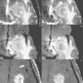
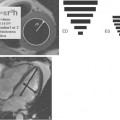
Suppressing the signal from fat (adipose tissue) in MR imaging is often very useful for improved lesion detection and delineation, as well as lesion characterization. The most common technique is spectral fat saturation (often referred to as “fat sat”). Axial postcontrast T1-weighted images of the lumbar spine at 1.5 T are presented without (Fig. 55.1A) and with (Fig. 55.1B) spectral fat saturation. Depicted is a primary bone tumor (an osteoblastoma) involving the posterior elements of the L5 vertebra. By suppression of the fat signal, abnormal contrast enhancement of the lesion, extending into the posterior paraspinal soft tissues, is much more evident. Fat saturation is commonly employed for postcontrast imaging of the soft tissues of the head and neck, spine, abdomen, and pelvis. Applications for fat saturation on precontrast scans are also common, notably in T2-weighted imaging of the breast and spine (the latter for improved visualization of vertebral body lesions and disk hydration) and for T1-weighted imaging of the pancreas.
Stay updated, free articles. Join our Telegram channel

Full access? Get Clinical Tree


