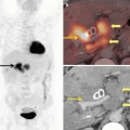
Fig. 5.1
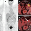
Poor FDG concentrating cholangiocarcinomas. Axial CT and fused PET/CT images shows a diffusely infiltrative type of cholangiocarcinoma showing low-grade FDG uptake (arrows in a, b). Axial CT and fused PET/CT images show extremely poor concentration of FDG in a diffuse mucinous type of cholangiocarcinoma (arrows in c, d)

Fig. 5.2
Poor FDG concentrating GB neck and cystic duct malignancy. Coronal MIP and fused PET/CT images (arrows in a, c) do not show significant FDG concentration in a stricturous lesion involving the neck of the GB and the cystic duct (arrow in b). Surgical resection revealed a mucin-producing adenocarcinoma
Cystic neoplasms of the pancreas include serous cystadenoma, mucinous cystadenoma and intraductal pancreatic neoplasms (IPMN). IMPNs are mucin-producing neoplasms that can be benign or invasive carcinomas, and FDG PET very often is used to differentiate between them [4]. Mucinous nature of these tumours causes poor avidity of FDG on PET studies (Fig. 5.3) more so in patients with a tiny malignant focus.


Fig. 5.3
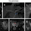
Poor FDG concentration in intraductal papillary mucinous neoplasm of pancreas. Axial CT scan shows a lobulated cystic mass arising from the pancreatic body (arrow in a) causing atrophy of the distal pancreatic body and tail (arrowhead in a, c). Axial PET and fused FDG PET/CT reveal no FDG uptake in the lesion. Histopathology after surgical resection showed IPMN with invasive features
5.3 Inflammatory Pathology Mimicking Malignancy
Mass-forming pancreatitis (MFP) resembles pancreatic adenocarcinoma on CT scan, and differentiating one from the other can be challenging. FDG PET/CT has been found to be better in distinguishing MFP from pancreatic adenocarcinoma by virtue of lower tracer uptake in the inflammatory lesion [5, 6]. However, certain cases of MFP can show high FDG avidity due to the inflammatory process and can simulate malignancy (Fig. 5.4).


Fig. 5.4
FDG avid mass forming pancreatitis. Coronal MIP, axial PET and fused PET/CT show increased FDG uptake (arrows in a, b, d) in a soft tissue mass seen in the pancreatic head (arrow in c). Biopsy revealed pancreatitis. FDG PET can be false positive in mass forming pancreatitis
Gall bladder wall thickening can be because of benign causes like inflammation and adenomyosis or due to malignancy. FDG PET can be falsely positive in cases of cholecystitis, and differentiation from malignancy can be difficult [7, 8]. Mass-like or protuberant lesions are more likely to be due to malignancy. Diffuse and uniform FDG avid wall thickening is usually due to inflammation (Fig. 5.5), though imaging features can overlap.


Fig. 5.5
FDG avid cholecystitis. Coronal PET and fusion PET/CT images (arrowheads in a, c) in a biopsy-proven case of cholangiocarcinoma show a FDG avid soft tissue lesion obstructing the common bile duct. FDG uptake along the thickened wall of the gall bladder (arrows in a–c) is due to cholecystitis and can be erroneously diagnosed as second malignancy
5.4 Pitfalls Due to Treatment-Related Changes and Complications
Significant proportion of gall bladder cancers are diagnosed incidentally from the surgical specimen after elective cholecystectomy is performed for symptomatic calculus disease. Staging PET/CT studies then performed often show FDG uptake in the operated bed of the gall bladder fossa which is due to postoperative inflammation associated with normal healing [2, 9]. This false-positive FDG uptake can persist for several weeks after surgery and mimic disease leading to futile surgical explorations to remove residual disease (Fig. 5.6).


Fig. 5.6


False-positive PET/CT after recent cholecystectomy. Coronal MIP, axial PET and fusion PET/CT images show intense FDG uptake in the GB fossa (arrows in a, b, d) in a patient with recent history (4 weeks) of cholecystectomy for symptomatic gall stones. Persistent FDG uptake is due to postoperative inflammation and can be confused with residual disease. No obvious mass lesion is seen in the GB fossa on axial CT image (arrowhead in c)
Stay updated, free articles. Join our Telegram channel

Full access? Get Clinical Tree


