(Left) Coronal graphic shows the normal course of the femoral nerve relative to the psoas muscle and inguinal ligament . The femoral nerve produces multiple peripheral branches to the anterior thigh muscles.
(Right) Coronal STIR MR (femoral neuropathy postsurgical herniorrhaphy) depicts marked enlargement, T2 hyperintensity of the right femoral nerve , with abrupt transition at the right groin. In this case, the femoral nerve was accidentally ligated during herniorrhaphy.
(Left) Axial STIR MR (femoral neuropathy postsurgical herniorrhaphy) through the pelvis confirms that the right femoral nerve in the iliopsoas groove is markedly enlarged with discrete T2 hyperintense fascicles (compared to the normal left femoral nerve ).
(Right) Axial CECT (severe hemophilia) depicts large left iliacus and psoas hematomas. Femoral neuropathy occurs from compression of the adjacent femoral nerve, which runs along the psoas muscle and iliopsoas groove.
 relative to the psoas muscle
  and inguinal ligament
  . The femoral nerve produces multiple peripheral branches to the anterior thigh muscles.
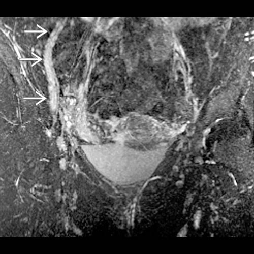
 , with abrupt transition at the right groin. In this case, the femoral nerve was accidentally ligated during herniorrhaphy.
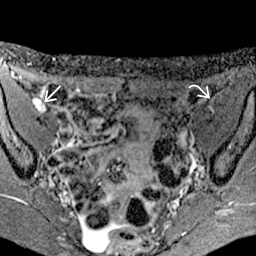
 in the iliopsoas groove is markedly enlarged with discrete T2 hyperintense fascicles (compared to the normal left femoral nerve
  ).
).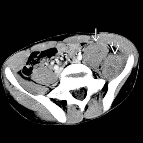
 and psoas
  hematomas. Femoral neuropathy occurs from compression of the adjacent femoral nerve, which runs along the psoas muscle and iliopsoas groove.


