Fig. 29.1
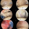
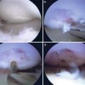
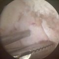
After cam lesions have been resected arthroscopically, adequacy of the resection is checked via direct arthroscopic visualization on dynamic examination. Hip is flexed to 90° in neutral rotation
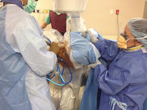
Fig. 29.2
Afterward, hip is rotated internally and externally to confirm no areas of impingement remain
Rehabilitation
An orthotic hip brace set for 0–90° flexion is utilized postoperatively while awake for 2 weeks, with formal physical therapy beginning on the first postoperative day. In addition, patient is instructed on the use of rotation precaution hip boots and pillow during sleep for 10 days to prevent inadvertent external rotation. Toe-touch weight bearing with crutches is implemented for 3 weeks after a formal femoral osteochondroplasty is performed in order to protect against torsion at the site of resection and femoral neck fracture. Protocol is modified for concomitant procedures such as microfracture by toe-touch weight bearing for 8 weeks, along with continuous passive motion for 6 h daily, lasting 4–6 weeks [20].
Case 1: Classic FAI in the Middle-Aged Working Male
History/Exam
A 49-year-old male, who works as a production supervisor, presented to the orthopedic sports clinic reporting 3 months of left hip pain when ascending stairs and doing computer work, requiring prolonged sitting. His pain measures 5/10 on VAS. He also reports pain with driving and getting into and out of his car. He has a history of right hip arthroscopy for FAI 2 years ago, for which he underwent labral debridement and femoroplasty for large cam lesion. His symptoms improved greater than 80 %. He states that pain is similar in nature to his contralateral hip prior to surgery, and it is preventing him from doing yard work; it requires him to bend down.
His physical examination revealed a positive flexion impingement test and pain with circumduction of the hip. It also demonstrated FABER asymmetry and weakness in hip musculature manifested by positive step-down and bridge tests. Passive range of motion was limited to flexion of 100°, IR 20°, ER 40°, abduction of 45°, and adduction of 20°. Due to the fact that he had failed extensive nonsurgical options and was unable to perform his ADLs, decision was made to proceed with imaging and surgical intervention. He also received 90 % effective, but temporary relief from an intra-articular cortisone injection lasting only a few weeks.
Imaging
Radiographs in Figs. 29.3 and 29.4 reveal extensive asphericity of his femoral head with significant cam lesion. His head–neck offset was significantly diminished and a minimal cross over sign. Lateral center-edge angle was measured at 24° with minimal joint space narrowing. Magnetic resonance imaging with contrast arthrography exhibited a manifestation of impingement via herniation pit on the lateral aspect of the femoral neck on the coronal images (Fig. 29.5). The axial images made it evident that the patient had a labral tear extending from anterior to superior (Fig. 29.6). Some mild adjacent chondral shearing was evident. Significant asphericity was again noted with decreased head–neck offset. Alpha angle was high and evidence of impingement was noted on the lateral aspect of head–neck junction.
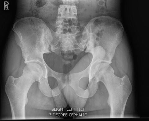
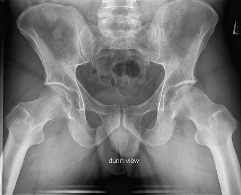
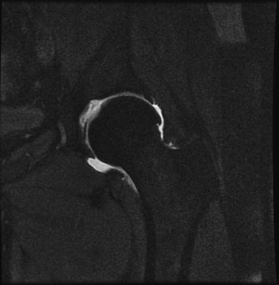
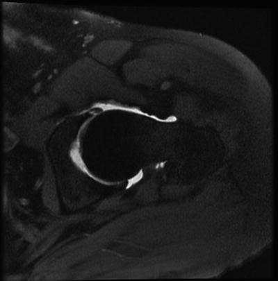

Fig. 29.3
AP pelvis demonstrates anterolateral cam lesion of left hip and loss of asphericity manifested by decrease in femoral head–neck offset when compared to contralateral hip

Fig. 29.4
Dunn view radiographs confirm this loss of femoral head–neck offset when compared to contralateral hip which has undergone previous hip arthroscopy, femoroplasty

Fig. 29.5
Coronal T2 MRA demonstrates anterolateral cam lesion with herniation pit manifested by high, bright fluid signal on lateral aspect of the head–neck junction

Fig. 29.6
Axial T2 MRA demonstrates same anterior cam lesion with adjacent anterior labral tear suggested by high, bright fluid signal at the labral–chondral junction
Arthroscopy
Patient was taken to the operating room for hip arthroscopy via technique stated above. After capsulotomy and labral repair were performed under distraction, arthroscopy of the peripheral compartment with the hip reduced revealed significant cam lesion on the anterolateral aspect of the head–neck junction seen in Fig. 29.7. Areas of impingement were noted and quantified in this area on arthroscopic impingement test. Thus, cam lesion was removed utilizing a 5-mm-round burr while viewing from the anterolateral portal and working through the mid-anterior portal (Fig. 29.8). A critical step in fully appreciating and adequately resecting the entire lesion is to switch the viewing and working portals in order to obtain a different vantage point. Figure 29.9 demonstrates this and allows complete resection of the lateral gutter. Cam resection measured 5-mm deep and 35-mm wide from a medial 6 o’clock position to a lateral 12 o’clock position. Periodic dynamic examination was performed to indicate sufficient resection with no impingement.
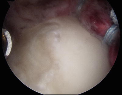
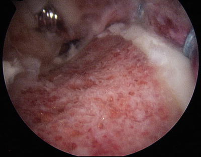
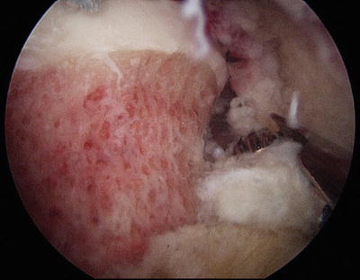

Fig. 29.7
Anterolateral cam lesion apparent during visualization of peripheral compartment via standard anterolateral portal after capsulotomy and labral repair seen in the background

Fig. 29.8
Combination of electrocautery and high-speed burr is utilized to resect the cam lesion to reestablish the femoral head–neck offset and relieve source of impingement

Fig. 29.9
Critical step in relieving entire cam lesion involves moving arthroscope to the distal anterior portal in order to adequately visualize and resect the lateral-most aspect of the lesion
After completion of femoroplasty, hip flexion is removed in neutral rotation and “T-capsulotomy” closure is performed with one nonabsorbable high-strength suture passed with a 90° suture-passing device and tied in a simple fashion (Figs. 29.10, 29.11, and 29.12).
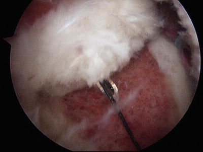
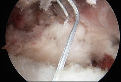
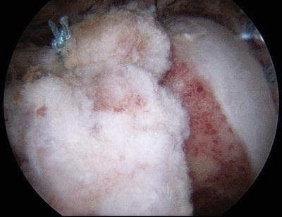

Fig. 29.10
A 90° suture-passing device is used via cannula in the mid-anterior portal to pass a nonabsorbable high-strength suture, aiding to close “T-capsulotomy”

Fig. 29.11
Nonabsorbable high-strength suture visible passed through each limb of the “T-capsulotomy,” exiting cannula via the mid-anterior portal

Fig. 29.12
One nonabsorbable high-strength suture tied in a simple fashion demonstrating closure of “T-capsulotomy” and good coverage of femoral neck and femoroplasty region
Discussion
The clinical history of reduced hip motion and increasing pain with deep-flexion activity coupled with prior success with contralateral side correction indicated imaging with MRI for this case. The MRI signs of articular chondral and labrum tearing in the anterior superior regions of the hip along with the femoral head asphericity were pertinent positives. Pertinent negatives included absence of severe subchondral cystic change and bone edema within the femoral head or acetabulum. The intraoperative decision making describes the importance of correlating preoperative imaging and exam findings with arthroscopic exam to adequately correct impingement.
Case 2: Atypical FAI in the Younger Male Athlete
History/Exam
A 17-year-old high school baseball player presented with bilateral hip pain, L>R, for the last 5 years that worsened with sports activity and twisting motions. He rated the pain as a 5/10 on a daily basis on the VAS. He had recently stopped participating in baseball due to his hip pain. The only relief from pain was rest from activities and anti-inflammatory medication.
On physical examination, there was no tenderness to palpation or crepitus. Both flexion impingement and posterior rim impingement tests were markedly positive. He also demonstrated a positive posterior rim impingement test. There was pain with circumduction of the hip. Patient also demonstrated weakness in hip musculature manifested by positive step-down and bridge tests. Patient’s passive range of motion was limited to flexion of 110°, IR 30°, ER 40°, abduction of 45°, and adduction of 20°. These symptoms with prolonged duration and recent regression in his chosen sport facilitated the decision to proceed with imaging studies and subsequent intervention.
Imaging
Radiographs demonstrated asphericity of the femoral head in bilateral hips. When examining the left hip, it was evident in Fig. 29.13 on the frog leg radiograph that there was a large posterior cam lesion in addition to the standard anterolateral lesion causing classic FAI. The lateral center-edge angle was measured at 35° with maintained joint space [21]. Magnetic resonance imaging with contrast arthrography confirmed the presence of anterior and posterior cam lesions. It also suggested a large labral tear manifested by significant contrast visualized medial to the labrum (Figs. 29.14 and 29.15). Enhancement of the tear is secondary to the use of contrast to improve sensitivity and specificity.
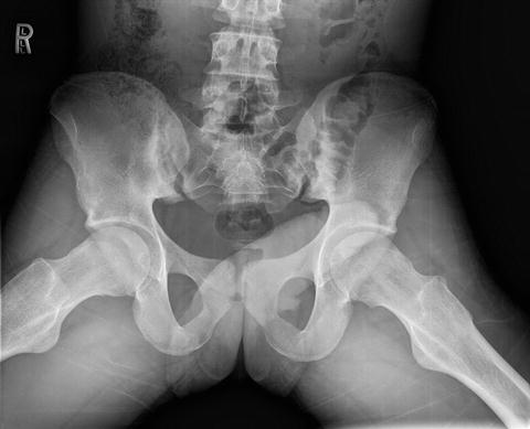
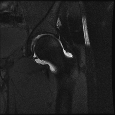
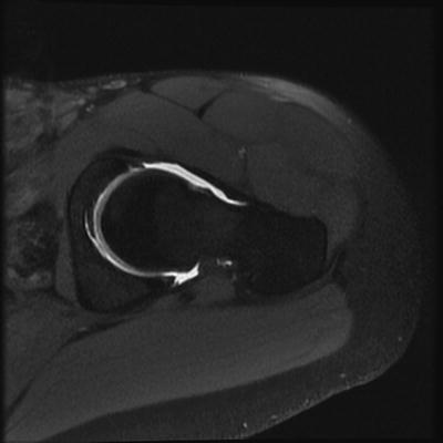


Fig. 29.13
Frog leg radiograph demonstrates bilateral anterior cam lesion with suspicion of posterior cam lesion and impingement in the left hip

Fig. 29.14
Coronal T2 MRA demonstrates asphericity of femoral head with evidence of a large anterior cam lesion and loss of femoral head–neck offset. A labral tear is suggested by a bright fluid signal medial to the labral tissue

Fig. 29.15
Axial T2 MRA identifies anterior and posterior cam lesions with evidence of a labral tear suggested by a bright fluid signal medial to the labrum
Stay updated, free articles. Join our Telegram channel

Full access? Get Clinical Tree


